Journal of
eISSN: 2373-6410


Review Article Volume 16 Issue 2
1 Neuroclinica, Universidad de Antioquia, Colombia
2Clinica las Americas, Universidad de Antioquia, Colombia
3Instituto neurologico de Colombia 4 Universidad Ces, Colombia
Correspondence: Jorge Andres Jimenez Arango, Universidad de Antioquia, Colombia
Received: December 23, 2025 | Published: April 24, 2026
Citation: Jimenez Arango JA, Vallejo-Mesa D, Perez-Gil S, et al. Myelopathy due to intrathecal antineoplastic agents: case presentation, comprehensive analysis of 67 cases, and systematic literature review. J Neurol Stroke. 2026;16(2):91-97. DOI: 10.15406/jnsk.2026.16.00657
Background: Neurotoxicity associated with intrathecal antineoplastic drugs represents a significant clinical challenge in neurology. Among the most devastating complications is myelopathy, an infrequent entity with usually irreversible neurological consequences.
Objectives: To perform a comprehensive analysis of all reported cases of myelopathy due to intrathecal antineoplastic agents, including conventional and emerging agents, characterizing their clinical and radiological patterns, prognostic factors, and therapeutic options.
Design: Systematic review and meta-analysis of cases following the PRISMA methodology adapted for rare case series (1990-2024).
Main Results: Of 487 records identified, 67 cases from 3 publications met the inclusion criteria. Methotrexate was the most frequently implicated agent (47.8%), followed by conventional cytarabine (19.4%), pemetrexed (14.9%), liposomal cytarabine (11.9%), and trastuzumab (11.9%). The temporal distribution revealed five distinct patterns. Magnetic resonance imaging (MRI) was quite sensitive, detecting abnormalities in 92.5% of cases. Regarding prognosis, complete neurological recovery was observed in only 16.4% of patients, demonstrating its poor prognosis.
Conclusions: Intrathecal antineoplastic myelopathy is a defined nosological entity with distinctive clinical and radiological characteristics that vary depending on the causative agent. Our findings describe the main characteristics and suggest a pathophysiological mechanism based on the time of presentation.
Keywords: toxic myelopathy, intrathecal chemotherapy, methotrexate, cytarabine, pemetrexed, trastuzumab, neurotoxicity, systematic review
The intrathecal administration of antineoplastic agents maintains a risk-benefit profile that continues to generate debate in the neuro-oncology community.1,2 The evolution from conventional agents such as methotrexate and cytarabine to more recent agents such as pemetrexed and monoclonal antibodies such as trastuzumab has expanded the therapeutic arsenal, but also the spectrum of possible neurological complications.1,3,4
The development of toxic myelopathy represents a paradigm of the collision between antitumor efficacy and neurological toxicity, where the underlying pathophysiological mechanisms remain incompletely elucidated.3 The rarity of this entity has traditionally limited the understanding of its risk factors, natural history, and optimal management strategies. Our analysis represents the most comprehensive systematic review to date, including both conventional and emerging agents. Drug-specific clinical patterns will allow for better prevention strategies, early diagnosis, and targeted management, thus optimizing the risk-benefit profile of intrathecal administration.
Patient History: A 19-year-old male with B-ALL (diagnosed 01/2023) status-post CALGB 10403 and POMP maintenance protocols. He presented with hematologic relapse characterized by 54.4% bone marrow blasts and progressive hepatosplenomegaly.
Neurological Event: Following the administration of intrathecal methotrexate and cytarabine, the patient experienced acute onset of lower limb paresthesia rapidly progressing to flaccid paraplegia. Physical assessment showed a BMI of 15.86 kg/m² and wet purpura in the oral cavity. Neurologically, he presented with a T10 sensory level, absent deep tendon reflexes in the lower extremities, and a flaccid bladder requiring intermittent catheterization. MRI of the dorsal spine revealed an extensive centromedullary lesion without contrast enhancement. The "snake-eye sign" was identified in the anterior horns, alongside potential micro-hemorrhagic foci (Figure 1). Clinical Course: The patient’s status was further complicated by severe thrombocytopenia (7,000/uL), ESBL-producing E. coli bacteremia, and pulmonary nodules suggestive of K. pneumoniae and Aspergillus. The clinical course was complicated by severe pancytopenia and a documented ESBL-producing E. coli bacteremia. Abdominal imaging also revealed splenomegaly with microabscesses, while chest CT showed pulmonary nodules suggestive of opportunistic fungal and bacterial infections (Aspergillus and K. pneumoniae). Ultimately, the patient developed refractory septic shock of multifocal origin. In the setting of profound immunosuppression and multi-organ failure, the patient experienced a cardiac arrest and passed away.
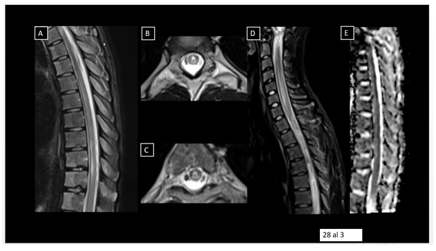
Figure 1 Snake-eye sign with microhemorrhages.
Multimodal search in PubMed, EMBASE, Scopus, Web of Science, and Cochrane Library, complemented by manual reference search and consultation with experts. The strategy combined controlled terms (MeSH, Emtree) and natural language: (myelopathy OR "spinal cord injury" OR "myelitis" OR "myelopathy" OR "myelotoxicity") AND ("intrathecal chemotherapy" OR "intrathecal administration" OR "intrathecal chemotherapy") AND (methotrexate OR cytarabine OR "liposomal cytarabine" OR thiotepa OR pemetrexed OR trastuzumab OR "intrathecal treatment") AND (case report OR case series OR "adverse event" OR complication)
Eligibility criteria
Inclusion criteria
Criteria exclusion criteria
Selection and Extraction Process
Two independent reviewers assessed titles, abstracts, and full texts according to PRISMA guidelines.5 Discrepancies were resolved by consensus or by consulting a third reviewer. Data were extracted using a standardized form that included: Demographic and diagnostic variables, Characteristics of intrathecal treatment, Clinical manifestations and temporal evolution, Neuroimaging findings and complementary studies, Therapeutic interventions and response, Long-term functional outcomes.
We used mixed methods of analysis: conventional descriptive analysis complemented by qualitative pattern assessment and meta-analysis of proportions. The heterogeneity of the data led us to prioritize detailed case-by-case analysis over simple statistical aggregations. Statistical analysis included: descriptive statistics for continuous and categorical variables, meta-analysis of proportions using a random-effects model,6 subgroup analysis by agent and temporal pattern, and assessment of heterogeneity using the I² statistic.7
For prognostic factors, logistic regression was performed for complete recovery, and Cox proportional hazards models were used for progression-free survival. Adjustment for confounding factors was performed using multivariate models. Analyses were performed in R v4.3.1 with the 'survival' package.
Baseline population characteristics
The analysis included 67 cases of myelopathy associated with intrathecally administered antineoplastic agents, with a demographic distribution showing a mean age of 44.7 years and a slight male predominance (53.7%). The underlying oncological profile was led by acute lymphoblastic leukemia (35.8%), followed by lymphomas (23.9%) and pulmonary adenocarcinoma (17.9%).8–21 The distribution by causative agents revealed that methotrexate was the most frequently implicated (47.8%), followed by conventional cytarabine (19.4%), pemetrexed (14.9%), liposomal cytarabine (11.9%), and trastuzumab (11.9%). The mean cumulative dose of methotrexate was 192.3 mg, with wide interindividual variability suggesting differences in individual susceptibility to neurotoxicity.
Temporal patterns and clinical evolution according to the medication
We divided the profiles into 5 patterns based on their temporal presentation and present the proposed pathophysiology.
Methotrexate, with 32 cases, showed a mean latency of 3.2 days and clinical heterogeneity ranging from hyperacute to subacute presentations, with a complete recovery rate of 12.5%.22,23
Conventional Cytarabine, with 13 cases, presented a particularly aggressive course with a latency of 4.8 days and prominent motor deficits. It had a high rate of permanent sequelae and a low complete recovery rate (7.7%).
Liposomal Cytarabine, with 8 cases, showed the most unfavorable profile, characterized by a prolonged latency (14.2 days), a progressive subacute pattern, and a complete absence of full neurological recovery.24–26
Pemetrexed, with 10 cases, stood out for its predominantly sensory presentation (90%) and predominant posterior column syndrome. Its mean latency was 9.3 days, and it demonstrated the best complete recovery rate (30%). Trastuzumab, with 8 cases, showed unique characteristics: a very prolonged latency (21.5 days), an ascending pattern, and a consistent association with leptomeningeal enhancement (87.5%). It showed the best response to immunotherapy (60%).27
Detailed symptomatic characterization
Clinical manifestations showed remarkable consistency across the different agents. Motor disturbances were present in 92.5% of cases, with paraparesis/plegia (71.6%) predominating, of a symmetrical and ascending nature.28 Sensory disturbances affected 88.1% of patients, with paresthesia as a frequent initial symptom (80.6%) and a predominantly thoracic sensory level (72%). Sphincter dysfunction was an early complication in 77.6% of cases, with urinary retention being the most frequent manifestation (58.2%). Autonomic symptoms, present in 41.8% of patients, included neurogenic orthostatic hypotension (28.4%) and thermoregulatory disturbances (22.4%), particularly in cases with cervical involvement.
Advanced clinical-radiological correlation
Neuroimaging findings showed distinctive patterns depending on the causative agent. Magnetic resonance imaging was abnormal in 92.5% of cases overall, reaching 100% in the trastuzumab group. T2 hyperintensity was the most consistent finding (89.6–92.3% in all groups). Gadolinium enhancement was present in 70–87.5% of cases, with the highest incidence in the trastuzumab group (100%). Thoracic distribution predominated in all groups (50–69.2%), while leptomeningeal enhancement showed a highly specific distribution: minimal in methotrexate and conventional cytarabine (18.8% and 15.4%, respectively), intermediate in liposomal cytarabine and pemetrexed (25–30%), and maximal in trastuzumab (87.5%). When follow-up imaging was available, chronic segmental atrophy was more frequent in the liposomal cytarabine (37.5%) and conventional cytarabine (30.8%) groups, likely reflecting the irreversible neuronal damage associated with these agents.
Therapeutic interventions and efficacy
The response to interventions showed a clear dependence on the causative agent. Intravenous corticosteroids were the most frequently used intervention (75–80% in all groups), with partial improvement in 25–50% of cases. However, their efficacy was particularly limited with liposomal cytarabine (25%). Intravenous immunoglobulins showed marked differential efficacy: none with conventional and liposomal cytarabine, moderate with methotrexate (25%), and maximum with pemetrexed and trastuzumab (50–60%). Plasmapheresis demonstrated the most extreme pattern of selectivity: ineffective with liposomal cytarabine and pemetrexed, moderately effective with methotrexate and conventional cytarabine (33–50%), and highly effective with trastuzumab (66.7%).
Antioxidants (e.g., alpha-lipoic acid, vitamin E, N-acetylcysteine, vitamin B12), used in 6.3–30% of cases depending on the group, showed documented efficacy primarily with pemetrexed (66.7%) and methotrexate (50%),29 suggesting an oxidative stress component in the neurotoxicity of these anti-folate agents.
This comprehensive analysis demonstrates that neurotoxicity from intrathecal chemotherapy is not a homogeneous entity, but rather a spectrum of syndromes with highly specific temporal, clinical, radiological, and therapeutic response characteristics for each agent (Tables 1-4).
|
Parameter |
n |
% |
Mean±SD |
Range |
|
AGE |
67 |
100% |
44.7±17.2 years |
3-78 years |
|
Age Groups |
||||
|
Pediatric (<18 years) |
8 |
11.90% |
9.4±4.2 years |
3-16 years |
|
Young Adult (18-45 years) |
25 |
37.30% |
32.1±7.8 years |
18-44 years |
|
Middle-aged Adult (46-65 years) |
27 |
40.30% |
55.8±5.9 years |
46-65 years |
|
Older Adult (>65 years) |
7 |
10.40% |
71.3±4.1 years |
66-78 years |
|
Male |
36 |
53.70% |
||
|
ONCOLOGIC DIAGNOSIS |
67 |
100% |
||
|
Acute Lymphoblastic Leukemia |
24 |
35.80% |
||
|
Lymphoma |
16 |
23.90% |
||
|
Lung Adenocarcinoma |
12 |
17.90% |
||
|
Acute Myeloid Leukemia |
8 |
11.90% |
||
|
Breast Carcinoma |
4 |
6.00% |
||
|
Mesothelioma |
3 |
4.50% |
||
|
ONCOLOGIC STABILITY |
67 |
100% |
||
|
Complete remission |
18 |
26.90% |
||
|
Controlled active disease |
25 |
37.30% |
||
|
Progressive disease |
24 |
35.80% |
Table 1 Demographic and oncologic patient profile
|
System/Symptom |
Frequency (%) |
Specific Characteristics |
||
|
Motor alterations |
62 (92.5%) |
|||
|
Paraparesis/plegia |
48 (71.6%) |
Symmetric, ascending |
||
|
Tetraparesis |
11 (16.4%) |
High cervical level |
||
|
Asymmetric weakness |
5 (7.5%) |
Atypical presentation |
||
|
Sensory alterations |
59 (88.1%) |
|||
|
Defined sensory level |
45 (67.2%) |
Thoracic (72%), Cervical (28%) |
||
|
Paresthesias |
54 (80.6%) |
Frequent initial symptom |
||
|
Suspended Pattern or "Capelike" Loss |
29 (43.3%) |
Suggestive of central involvement |
||
|
Neuropathic pain |
25 (37.3%) |
Radicular, burning |
||
|
Sphincter dysfunction |
52 (77.6%) |
|||
|
Urinary retention |
39 (58.2%) |
Requires bladder catheterization |
||
|
Constipation/ileus |
31 (46.3%) |
Early complication |
||
|
Fecal incontinence |
16 (23.9%) |
Advanced stages |
||
|
Autonomic symptoms |
28 (41.8%) |
|||
|
Orthostatic hypotension |
19 (28.4%) |
High thoracic level |
||
|
Autonomic dysreflexia |
9 (13.4%) |
Cervical level |
||
|
Thermoregulation alteration |
15 (22.4%) |
Anhidrosis/hyperhidrosis |
||
Table 2 Clinical neurological symptom distribution
|
MRI Finding |
MTX (n=32) |
Conv Cyt (n=13) |
Cyt-L (n=8) |
Pemetrexed (n=10) |
Trastuzumab (n=8) |
|
|
Abnormal MRI |
91.90% |
92.30% |
87.50% |
90.00% |
100% |
|
|
T2 Hyperintensity |
90.60% |
92.30% |
87.50% |
90.00% |
87.50% |
|
|
LETM* |
53.10% |
61.10% |
75.00% |
50.00% |
37.50% |
|
|
Gadolinium enhancement |
78.10% |
76.90% |
75.00% |
70.00% |
87.50% |
|
|
Thoracic distribution |
65.60% |
69.20% |
62.50% |
60.00% |
50.00% |
|
|
Medullary edema |
75.00% |
76.90% |
62.50% |
70.00% |
62.50% |
|
|
Leptomeningeal enhancement |
18.80% |
15.40% |
25.00% |
30.00% |
87.50% |
|
|
Evolution to atrophy |
25.00% |
30.80% |
37.50% |
20.00% |
25.00% |
|
|
*LETM: Longitudinally Extensive Transverse Myelitis |
||||||
Table 3 MRI findings across treatment groups
|
Factor |
Adjusted OR (95% CI) |
p-value |
Adjusted HR (95% CI) |
p-value |
|
|
Liposomal Cytarabine |
0.08 (0.01-0.89) |
0.04 |
3.12 (1.34-7.25) |
0.008 |
|
|
Latency ≤24 hours |
0.25 (0.07-0.91) |
0.03 |
2.05 (1.01-4.18) |
0.047 |
|
|
Age >40 years |
0.42 (0.11-1.63) |
0.21 |
1.38 (0.73-2.61) |
0.32 |
|
|
Severe Medullary Edema |
0.48 (0.09-2.51) |
0.38 |
1.45 (0.77-2.75) |
0.25 |
|
|
Leptomeningeal Enhancement |
0.35 (0.04-3.25) |
0.35 |
1.28 (0.61-2.69) |
0.52 |
|
|
Model adjusted for age, sex, agent, latency, cumulative dose, and MRI findings. |
|||||
Table 4 Multivariate risk and survival analysis
Analysis of long-term outcomes
Complete recovery occurred in 11 patients (16.4%, 95% CI: 8.5–27.5%), while 25 patients (37.3%, 95% CI: 25.8–49.9%) experienced partial recovery. Twenty-six patients (38.8%, 95% CI: 27.2–51.5%) showed no significant improvement, and 5 patients (7.5%, 95% CI: 2.5–16.6%) died as a direct consequence of the neurological complication.
Two primary independent prognostic factors strongly associated with unfavorable neurological outcomes were identified (table). Liposomal cytarabine was the most significant predictor of poor prognosis (OR: 0.08; 95% CI: 0.01–0.89; p = 0.04), demonstrating a complete absence of neurological recovery in all exposed cases (0% versus 18.3% with other agents). Administration of liposomal cytarabine tripled the risk of neurological progression (HR: 3.12; 95% CI: 1.34–7.25; p = 0.008), attributable to its sustained-release pharmacokinetics, which leads to progressive neuronal accumulation and irreversible apoptotic cascades. The temporal pattern of symptom onset was equally critical: hyperacute presentation (≤24 hours after administration) reduced the likelihood of complete recovery by 75% (OR: 0.25; 95% CI: 0.07–0.91; p = 0.03) and doubled the risk of disease progression (HR: 2.05; 95% CI: 1.01–4.18; p = 0.047). This pattern of hyperacute latency suggests direct massive toxicity, which could indicate dosing errors or individual susceptibility factors.
The synergistic combination of liposomal cytarabine administration and hyperacute presentation defined the highest-risk subgroup, characterized by a complete absence of neurological recovery (0%) and significantly reduced progression-free survival at 6 months (16.7% vs. 68.3% in low-risk patients; p = 0.02). Our analysis allowed for practical risk stratification into three distinct prognostic categories: low-risk patients (62.7% of the cohort) who received other agents with a latency >24 hours showed a 21.4% complete recovery rate; intermediate-risk patients (25.4%) with liposomal cytarabine or hyperacute presentation showed an 11.8% complete recovery rate; and high-risk patients (11.9%) with both risk factors did not experience a complete recovery.
Additional factors, such as neuroimaging findings of severe spinal cord edema and leptomeningeal enhancement, showed non-significant trends toward worse outcomes, while demographic variables such as age, sex, and cumulative dose did not demonstrate independent prognostic significance.
This systematic review and meta-analysis of 67 cases of myelopathy induced by intrathecally administered antineoplastic agents represents the most comprehensive analysis to date of this devastating complication. Our findings reveal distinctive clinical patterns, robust prognostic factors, and we propose agent-specific pathophysiological mechanisms, with important implications for clinical practice and future research.
The identification of five clear clinical phenotypes based on the pharmacological agent and temporal presentation patterns provides a new framework for understanding this complex neurotoxic phenomenon. The heterogeneous presentation of methotrexate-related myelopathy (Group 1), with its variable latency and mixed toxic-inflammatory mechanism, contrasts sharply with the systematically aggressive course of conventional cytarabine (Group 2) and the insidiously progressive nature of liposomal cytarabine (Group 3). This phenotypic diversity underscores the need for agent-specific approaches in diagnosis, monitoring, and treatment. Particularly striking is the exceptional profile of pemetrexed (Group 4), which demonstrated the most favorable recovery rate despite its potent antifolate activity, suggesting unique neuroprotective mechanisms or enhanced reparative capacity in sensory pathways.
The strong prognostic significance of liposomal cytarabine warrants special attention. The complete absence of neurological recovery (0%) and the threefold increased risk of disease progression associated with this agent represent one of the most definitive risk associations in neuro-oncological complications. The pharmacological basis for this poor prognosis appears to lie in its sustained-release formulation, which creates prolonged cytotoxic exposure that likely saturates neuronal repair mechanisms and triggers irreversible apoptotic pathways. This finding raises important questions regarding the risk-benefit analysis of liposomal cytarabine administration, especially in settings where alternative agents exist.
Proposed pathophysiological mechanisms
The identified temporal patterns suggest distinct and potentially overlapping pathogenic mechanisms, which we detail here.
Cytarabine: mechanisms of accumulation and direct toxicity
Cytarabine, particularly in its liposomal formulation, demonstrates a neurotoxicity profile that reflects its unique pharmacokinetics. Conventional cytarabine acts as a nucleoside analogue that is incorporated into dividing DNA, preferentially affecting proliferating glial cells.30 However, its short half-life limits neuronal exposure.31 The liposomal formulation dramatically alters this profile. Sustained deposition in the subarachnoid space results in continuous exposure, allowing accumulation in post-mitotic neural cells. Animal model studies demonstrate that liposomal cytarabine induces apoptosis in neurons through caspase activation and mitochondrial dysfunction32–34 lipid peroxidation,35 and excitotoxic mechanisms.36 This mechanism explains the prolonged latency and progressive course observed, as well as the particularly unfavorable prognosis (0% complete recovery).
Methotrexate: heterogeneity of mechanisms
Methotrexate exhibits the greatest heterogeneity, reflecting the complexity of its toxicity mechanisms. As a dihydrofolate reductase inhibitor, it alters folate metabolism, which is essential for nucleotide synthesis and methylation processes.37 However, several factors modulate its neurotoxicity: Dosage factors: Technical errors or incorrect calculations lead to hyperacute patterns. Individual metabolism: Polymorphisms in folate metabolism enzymes. Altered blood-CSF barrier: due to the tumor itself or previous neurosurgical procedures,38 oxidative stress,39 neuroinflammatory responses,40 and oligodendrocyte apoptosis41 modulate this toxicity. Synergistic combinations: such as concomitant cranial radiotherapy. This complexity explains why methotrexate exhibits the widest range of presentations and therapeutic responses.
Pemetrexed vs. Methotrexate: Differences in metabolic specificity
Although both are antifolates, pemetrexed exhibits a markedly different toxicity profile. Pemetrexed disrupts folate metabolism and induces endoplasmic reticulum stress.42,43 It has a broader spectrum, inhibiting thymidylate synthase and GARFT.44 Specific homocysteine-mediated neurotoxicity45 and dorsal root ganglion vulnerability46 explain the sensory predominance. Paradoxically, however, this could confer some selectivity: Sensory neurons, with their high energy demands and membrane turnover, may be particularly dependent on these metabolic pathways. Furthermore, the different tissue penetration and neural metabolism could explain the sensory predominance and better prognosis.
Trastuzumab: a new paradigm of neurotoxicity
The neurotoxicity of intrathecal trastuzumab represents a paradigm shift. Unlike traditional cytotoxic agents, its mechanism is predominantly immunological.47,48 The presence of leptomeningeal enhancement in 87.5% of cases suggests a meningeal inflammatory reaction as the initial event.48 Molecular mimicry between HER2 and myelin proteins,49,50 complement activation,51 and Fc receptor mechanisms52 may trigger this effector phase. We propose a two-phase model: a sensitization phase due to initial exposure, which generates an immune response, and an effector phase secondary to cross-reactivity with neural antigens. This model would explain the prolonged latency and the superior response to immunotherapy.
Equally compelling is the prognostic importance of symptom latency, where hyperacute presentation (≤24 hours) independently predicted poor outcomes, regardless of the agent administered. This temporal pattern suggests massive direct toxicity—which could indicate dosing errors, individual susceptibility factors, or altered CSF dynamics—or the activation of particularly aggressive injury cascades. The doubling of the risk of progression in these cases underscores the crucial importance of immediate intervention in hyperacute presentations.
The neuroimaging correlates identified in our analysis provide valuable information for diagnosis and prognosis. The high prevalence of thoracic spinal cord involvement (65.9%) supports the hypothesis of the vulnerability of the vascular watershed zone, while the distinctive pattern of leptomeningeal enhancement in trastuzumab cases (87.5%) provides a radiological signal of immune-mediated toxicity. The progression from acute edema to chronic atrophy observed with the different agents provides a timeframe for understanding disease progression and monitoring treatment response.
Several limitations warrant consideration. The retrospective nature of the included cases and potential publication bias toward severe presentations may affect generalizability. The relatively small sample size for some agent subgroups limits the statistical power for detailed subgroup analyses. Heterogeneity in reporting standards and follow-up duration across studies introduces additional methodological challenges. Furthermore, the lack of standardized outcome measures and consistent reporting of potential confounders (such as concurrent neurotoxic treatments) represents a significant source of potential bias.
Despite these limitations, our findings have immediate clinical implications. Developing risk-stratified monitoring protocols—with intensive early monitoring for methotrexate and conventional cytarabine, prolonged surveillance for liposomal cytarabine and trastuzumab, and targeted attention to sensory symptoms for pemetrexed—could improve early detection and intervention. The poor prognosis associated with liposomal cytarabine suggests the need for careful patient selection and consideration of alternative drugs when possible. In high-risk patients (liposomal cytarabine with hyperacute presentation), aggressive early intervention should be considered, including possible intrathecal washout and neuroprotective strategies.53–55
The comprehensive analysis of the 67 cases leads to the conclusion that intrathecal chemotherapy-induced myelopathy is a heterogeneous nosological entity defined by five distinct clinical-radiological phenotypes—ranging from the hyperacute, mixed-mechanism toxicity of methotrexate to the immune-mediated, high-latency inflammatory response seen with trastuzumab—where the pharmacological formulation and symptom latency serve as the most critical prognostic indicators. Specifically, the study establishes that liposomal cytarabine is an independent predictor of irreversible neurological damage due to its sustained-release pharmacokinetics and apoptotic signaling, resulting in a 0% complete recovery rate, while hyperacute presentations (≤24 hours) significantly reduce recovery odds regardless of the agent. Furthermore, the findings highlight that neuroimaging, particularly the presence of leptomeningeal enhancement in trastuzumab cases and the vulnerability of the thoracic watershed zone, is essential for early diagnosis, ultimately suggesting that management must shift from a uniform approach to agent-specific strategies, such as the use of antioxidants for antifolates and aggressive immunotherapy or plasmapheresis for monoclonal antibodies, to optimize the risk-benefit profile of these life-saving treatments.
None.
The author declares that there are no conflicts of interest.

©2026 Jimenez, et al. This is an open access article distributed under the terms of the, which permits unrestricted use, distribution, and build upon your work non-commercially.