Journal of
eISSN: 2373-6437


Review Article Volume 17 Issue 5
1Researcher without institution, Anesthesiologist at various hospitals, Brazil
2Professor, Department of Anesthesiology, Rohailkhand medical College and Hospital, India
3Hospital Santa Isabel and Santa Casa da Bahia, Brazil
4Consultant, Ganga Medical Centre and Hospital Pvt Ltd, Índia
5Anesthesiologist at the National Cancer Institute (INCA), Brazil
6Head of the Anesthesiology Service of the National Cancer Institute (INCA) Anesthesiologist, Responsible for the CET-SBA of the National Cancer Institute, Brazil
7Anesthesiologist of Hospital Anis Rassi, Brazil
8Graduated from Pontifical Catholic University of Goiás, Brazil, Fellowship in Integrative Medicine at the University of Arizona, USA
9Resident in Anesthesiology and pediatrics, USA
Correspondence: Dr. Luiz Eduardo Imbelloni, Researcher without institution, Anesthesiologist at various hospitals, Av. Epitácio Pessoa, 2356 - Apto 203, Lagoa 22411-072 – Rio de Janeiro, RJ, Brazil, Tel 55.11.99429-3637
Received: September 19, 2025 | Published: September 29, 2025
Citation: Imbelloni LE, Chandra R, Pitombo PF, et al. Anatomical and physiological factors for a lower incidence of post-dural puncture headache during thoracic spinal anesthesia. Narrative review. J Anesth Crit Care Open Acces. 2025;17(4):129‒134. DOI: 10.15406/jaccoa.2025.17.00634
Post-dural puncture headache (PDPH) is a side effect of the first spinal anesthesia and can also occur after an accidental dural puncture during epidural anesthesia. The risk of PDPH can be influenced by the size, shape, and orientation of the spinal needles, as well as the patient's posture. Leakage of cerebrospinal fluid (CSF) through the dura mater opening leads to traction on pain-sensitive structures, causing PDPH. There are various proposed mechanisms explaining how headaches are brought on by CSF hypotension. Spinal CSF dynamics are sensitive to varying respiratory performances. Since the beginning of the last century, subarachnoid blocks can be performed at any of the thoracic and lumbar spinal levels. Thoracic spinal anesthesia has been extensively studied in the 21st century, as have several new indications for spinal anesthesia. Thoracic spinal anesthesia (TSA) has been extensively studied in the 21st century, as have several new indications for spinal anesthesia. Several published articles on TSA demonstrate its safety, with a lack of neurological complications, particularly PDPH. Several mechanisms have been implicated in the lower incidence of thoracic puncture compared to lumbar puncture. This article shows several mechanisms for this lower incidence, and the low incidence after TSA compared with lumbar spinal anesthesia (LSA).
Keywords: Thoracic spinal anesthesia, post-dural puncture headache, cerebrospinal fluid.
Key Points
Meaning
Postdural puncture headache (PDPH) can occur because of diagnostic lumbar puncture, spinal anaesthesia, and accidental dural puncture during epidural anesthesia. Four studies on thoracic epidural anesthesia showed that in 1,071 patients,1 4,185 patients,2 1,240 patients3 and 113 patients4 there were 50 (0.75%) accidental perforations of the spinal dura mater with a large gauge needle, no neurological sequelae and no PDPH. The fact that there was no neurological injury led us to question whether there was anatomical protection or divine protection. And indeed, a magnetic resonance imaging (MRI) study in adult patients,5,6, and in children aged 0 to 13 years,7 showed a space between the spinal cord and the dura mater with protection for the needle tip.
Regarding the absence of neurological spinal cord injury, both studies demonstrated anatomical protection. However, since none of the four studies post accidental puncture during epidural anesthesia reported PDPH, this prompted us to investigate this fact: because accidental perfusion of the thoracic region with a large-bore needle does not result in this complication?
(Whether an accidental perfusion of the thoracic region with a large-bore needle does not result in this complication?)
After MRI studies, I began my studies to perform TSA in 300 patients, comparing Quincke and Whitacre needles. Paresthesias occurred in 20/300 (6.6%) of patients, with no statistical difference between the needle tip designs.7 All paresthesias were transient, and no neurological complications were observed in all patients during this study, and there were no reports of PDPH. From 2010 to 2025, eight studies on TSA have been conducted by me and my research group, with a total of 3,791 patients, and 4 (0.1%) headaches have been reported, all light (grade 1/3) easily treated with clinical methods and without the need to use a blood patch. All these articles will be included when the incidence of PDPH during TSA is addressed, and a survey of all published TSA. After more than 15 years of experience with TSA, I have found that the incidence of PDPH is significantly lower when the puncture is performed at the thoracic level compared to the lumbar region. Two articles were recently written addressing TSA with its indications, safety, anatomy and approach to the subarachnoid space in the thoracic region.8,9 Thus, this study aimed to determine whether there are any anatomical, physiological, and cerebrospinal fluid (CSF) related explanations.
Anatomy of the thoracic intervertebral space
The spinal canal is narrower than the cervical and lumbar regions, leaving less epidural and subarachnoid space volume. The spinal cord in the thoracic region occupies a larger proportion of the spinal canal, resulting in less free space between the spinal cord and the dura mater. Studied lesions of the human dural sac produced by different spinal needles and different bevel orientations. The dura mater has a thickness of around 400 μm, and it is formed by randomly distributed fibers, arranged around 80 concentric layers, known as dural laminas, while the arachnoid layer has a thickness of around 40 μm.10,11 The interlaminar spaces in the thoracic spine are narrow and more difficult to access with a needle due to the overlapping vertebral lamina, and the spinous processes of the thoracic vertebrae point downward.12 Intrathecal injections at mid-thoracic levels may have a minimum safe distance before the spinal needle contacts the spinal cord tissue.13 The spinal dura mater is a key component of the thoracic dura, and its anatomy plays a significant role in its function. The spinal dura mater is a tough fibrous membrane that protects and encloses the spinal cord. Understanding the biomechanics of the thorax is crucial, as it affects how the thoracic dura functions and can impact the closure of holes.14
Physiology of the thoracic intervertebral space
A smaller number of nerve roots are covered by an anesthetic within the subarachnoid space in TSA, providing anesthesia in the necessary surgical field dermatomes. Further, since there is less of a block of lower extremities, a larger portion of the body does not experience venous dilation, which may compensate for adverse effects on blood pressure intraoperatively.15
CSF formation and volume
CSF is essential for the mechanical and homeostatic protection of the central nervous system and the spinal column. CSF is produced primarily in the choroid plexus at a rate of approximately 0.35 mL/min and reabsorbed through the arachnoid villa, and a smaller part is formed by secretion of the ependyma and passage of interstitial fluid from the central nervous system (CNS) to the subarachnoid space.16 The total volume in adults is around 150 mL, with 20-30 mL in the ventricles, 80-100 mL in the cranial subarachnoid space, and 25-30 mL in the spinal subarachnoid space, with lumbar pressure of 5-15 cm H2O in the horizontal position and 40-50 cm H2O in the vertical position.16 PDPH is due to the loss of CSF through a persistent leak in the meninges.17 It has been shown experimentally that the loss of approximately 10% of total CSF volume predictably results in the development of typical PDPH symptoms, which resolve promptly with reconstitution of this deficit.
The influence of body position on CSF circulation
CSF plays an important role in providing structural and homeostatic support to the central nervous system.18 Evaluating 30 healthy volunteers in the upright and supine sitting positions with 0.6 T multiposition MRI (Fonar, New York, USA), CSF flow and spinal cord pulsation were visualized and quantified at the mid-axial level of C2 with contrast, showed that in upright posture, heart rate increased by 10%, and peak diastolic CSF flow decreased by 43% compared to the supine posture.19 The body position has significant effects on CSF flow in and out of the cranium, with more CSF oscillating in the supine compared to the upright position.19
CSF in the thoracic and lumbar subarachnoid space
CSF is present throughout the subarachnoid space, surrounding the spinal cord and brain. However, the distribution of CSF and the relationship between the spinal cord, nerve roots, and meninges vary between the thoracic and lumbar regions. In the thoracic region, the CSF volume is smaller, because the subarachnoid space is narrower around the spinal cord in this region, and the spinal cord occupies a greater proportion of the spinal canal, with less free space between the spinal cord and the dura mater.20 In the lumbar region, the volume of CSF is greater, and the spinal cord has already ended, with only the cauda equina existing, and the spinal canal is relatively wider, and the subarachnoid space contains a large amount of free CSF around the nerve roots.20
CSF volume in the thoracic space compared with the lumbar space
CSF circulation consists of two components, a net flow and a pulsatile flow. The pulsatile driving forces include cardiac vascular pulsation, respiration and muscular contraction. MR myelography (MRM) is an effective tool for detecting CSF leak in the spine in patients with spinal CSF leak syndrome.21 The thoracic region has a smaller subarachnoid space than the lumbar region, providing important space for the dispersion of intrathecal medications. Because it has a smaller CSF reservoir, it provides less loss, resulting in a lower incidence of PDPH. Furthermore, the spinal cord and roots are heavily surrounded by CSF and meninges, with less free mobility than the lumbar cauda equina. The smaller CSF volume in the thoracic region provides faster drug dissemination (short latency) and more restricted dispersion of local anesthetics injected into the subarachnoid space.
Influence of respiration and heart rate on CSF
Respiration-induced pressure changes represent a powerful driving force behind CSF dynamics. Eighteen subjects without known illness using contrast flow MRI, comparing forced thoracic versus abdominal breathing, showed that spinal CSF dynamics are sensitive to varying respiratory performances.22 Concluding that forced inspiration and expiration therefore lead to upward and downward CSF flow in the spinal canal. CSF has been thought to mainly follow cardiac-related oscillations as suggested by electrocardiogram-synchronized cine flow MRI.23 Through a mathematical model, it is suggested that the interaction between thoracic pressure and the cardiovascular system, particularly the central veins, has a greater influence on CSF pressure.24
Different characteristics of lumbar puncture and thoracic puncture
Several characteristics differ when performing a lumbar puncture, recommended by most anesthesiologists, compared to a thoracic puncture, which has seen significant development in the 21st century, as illustrated by two fundamental articles.8,9 The most important characteristics are: CSF volume, CSF pressure, epidural space, dura mater fibers, influence on respiration and heart on CSF (Table 1).
|
Characteristics |
Lumbar Spinal Anesthesia |
Thoracic Spinal Anesthesia |
|
CSF volume |
Greater amount in subarachnoid space |
Less amount in thoracic space |
|
CSF pressure |
Higher in a sitting position |
Lower reducing leakage |
|
Epidural space |
Wider, less positive pressure |
Narrower, more positive pressure |
|
Dura mater fibers |
Less resistant hole open longer |
More resistant hole closes faster |
|
Technique and needles |
Fine (25G, 26G, 27G, 29G) |
Fine (25G, 26G, 27G,29G) |
|
Incidence of PDPH |
Higher with thick needle |
Very low, rarely reported |
Table 1 Comparison between lumbar puncture and thoracic puncture
Incidence of PDPH after lumbar spinal anesthesia
Performed between the lumbar vertebrae (L2-L3, L3-L4, L4-L5), this is below the termination of the spinal cord (which ends around L1-L2 in adults), in the cauda equina region. PDPH is occasionally an inevitable side effect of neuraxial anesthesia, which can happen after spinal anesthesia or if an accidental dural puncture happens during epidural anesthesia. However, PDPH is more frequently caused by dural puncture during epidural anesthesia than by spinal anesthesia because spinal anesthesia uses small needles, in non-obstetric patients.25 PDPH is believed to be due to reduced brain pressure caused by CSF leakage. A decline in CSF pressure leads to traction on pain-sensitive parietal dura and intracranial structures, thereby causing subsequent headache in patients. When a pencil-point spinal needle is used, the risk of PDPH is reduced.26 The risk of PDPH can be influenced by the size, shape, and orientation of the spinal needles, as well as the patient's posture.27 The difference between the incidence of PDPH after LSA in non-obstetric patients in the different studies can be explained by the needle gauge and design, patient age, body mass index, history of chronic headache, puncture technique, direction of needle bevel and number of punctures.
Incidence of PDPH after thoracic spinal anesthesia
Performed between the thoracic vertebrae (e.g., T2-T11), at this level, the spinal cord is present and occupies most of the spinal canal. Patients are typically positioned either in the lateral recumbent or upright sitting posture. Spontaneous intracranial hypotension typically presents with a positional headache caused by downward displacement of the brain due to reduced buoyant support. It has long been accepted that PDPH results from a disruption of normal CSF homeostasis. CSF leakage from the dura, which leads to traction on pain-sensitive structures, is the cause of PDPH. The association between needle size and type of needle with the incidence of PDPH was described. However, despite a great deal of research and observational data, the pathophysiology of PDPH remains incompletely understood. In an article defining the role of thoracic spinal anesthesia in the 21st century, the authors do not address the incidence of PDPH after thoracic puncture.8 In another article evaluating the state of the art of Jonnesco's TSA to date, PDPH after thoracic puncture was not addressed.9 Performing a search in the various TSA publications to verify the incidence of PDPH in these publications correlating with the needles used (Table 2).
|
Ref |
Patients |
Needle |
PDPH |
|
15 |
20 |
Q=20 |
0 |
|
28 |
300 |
W=150 / Q=150 |
0 |
|
29 |
70 |
W=70 |
0 |
|
30 |
636 |
W=315 / Q=321 |
0 |
|
31 |
200 |
W=98 / Q=102 |
0 |
|
32 |
296 |
W=153 / Q=143 |
4 |
|
33 |
200 |
W=NR / Q=NR |
0 |
|
34 |
1406 |
W=697 / Q=709 |
0 |
|
35 |
505 |
W=NR / Q=NR |
0 |
|
36 |
674 |
W=332 / Q=341 |
0 |
|
37 |
40 |
Q=40 |
0 |
|
38 |
28 |
Q=28 |
0 |
|
39 |
2102 |
Q=2102 |
0 |
|
40 |
39 |
Q=39 |
0 |
|
41 |
78 |
Q=78 |
0 |
|
42 |
30 |
NR |
0 |
|
43 |
60 |
Q=60 |
0 |
|
44 |
50 |
Q=50 |
1 |
|
Total |
6734 |
|
5 (0.07%) |
Difference in incidence of PDPH lumbar and thoracic puncture
Start this topic with a question: Is there a difference in incidence of PDPH lumbar puncture and thoracic puncture for spinal anesthesia? The short answer is that there is a significant and well-established difference in the incidence of PDPH between lumbar and thoracic punctures. Since its inception by Bier,45 lumbar puncture has been considered the standard approach for spinal anesthesia and carries a well-known risk of PDPH. Thoracic puncture, pioneered by Jonnesco46 for spinal anesthesia, is extremely rare, used more in the 21st century and by a few authors, but with increasing use, making direct comparison in clinical practice difficult. PDPH is one of the complications following the first spinal anesthesia described by Bier and performed on his assistant and during accidental dural puncture. Several modifiable risk factors contribute to the development of headache after lumbar puncture, including needle size, needle design, direction of the bevel, and number of lumbar puncture attempts. In 5050 non-obstetric patients used 25G, 26G, 27G or 29G Quincke needles; 26G Atraucan or 27G Whitacre without introducer, the incidence was 0.8%.25 Regarding the caliber and tip design, the result was 25G Quincke 3%, 26G Quincke 1.4%, 27G Quincke 0.7%, 26G Atraucan 0.4%, 27G Whitacre 0.4% and 29G Quincke 0.3% (Table 3).25 The association between the gauge and type of needle tip with the incidence of PDPH was described with different types of needles, being greater with thicker needles (22G, 23G and 25G) and lower with thinner needles (26G, 27G and 29G) with the different types of Whitacre and Quincke tip.25
|
Caliber |
Needle |
Patients |
PDPH |
Incidence |
|
25G |
Quincke |
324 |
10 |
3% |
|
26G |
Quincke |
138 |
2 |
1.4% |
|
26G |
Atraucan |
220 |
1 |
0.4% |
|
27G |
Quincke |
3234 |
23 |
0.7% |
|
27G |
Whitacre |
210 |
1 |
0.4% |
|
29G |
Quincke |
924 |
3 |
0.3% |
|
Total |
|
5050 |
40 |
0.8% |
Table 3 Incidence of PDPH in an article published on LSA.25
Use of the tip design of fine-gauge needles in thoracic puncture
In the thoracic dura, fibers are denser and more compact, creating a tighter, less compliant sheath. When a spinal needle penetrates the thoracic dura, the longitudinal orientation makes the puncture track more likely to close, reducing CSF leakage compared to a lumber puncture. However, when thoracic puncture was used to perform TSA, it was shown in 6,734 patients from various publications punctured with Whitacre or Quincke needles that the incidence was only 0.07% (Table II). Several explanations were shown in this article. Direction of the spinal needle is seen to be that mid or lower thoracic punctures need a steep angle for penetration of the structures, and an angle of 45 degrees is often recommended.4 This angulation gives a less direct communication between the epidural and the dural rent leading to less PDPH.
Orientation of dura mater fibers in the thoracic and lumbar regions
The dura mater is of similar embryological origin to the fascial organ. It contains several fibroblasts which make the dura mater a flexible structure (Figure 1). The dura mater is the outermost layer of the meninges and plays a crucial role in protecting the spinal cord. The dura mater in the thoracic spine has a very organized fiber orientation, which is relevant to why PDPH is less common after thoracic puncture than after lumbar puncture. The collagen and elastic fibers in the thoracic dura run predominantly in a longitudinal (craniocaudal) direction, parallel to the spinal axis.47 These fibers are arranged in lamellae (layers), with most oriented longitudinally, but with some interlacing oblique and transverse fibers providing tensile strength.47
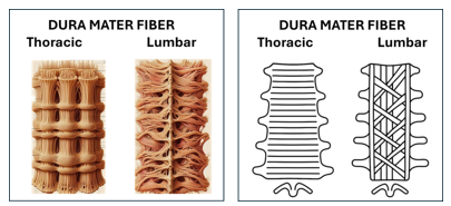
Figure 1 Fibers of the dura mater in the thoracic region and lumbar region and copilot design.
Why is thinner dura less prone to tearing?
The dura mater is the tough outermost membrane covering the brain and spinal cord, and its thickness can vary depending on the region of the spine.48,49 Thinner dura is generally less prone to tearing due to its greater elasticity, reduced resistance to penetration, lower internal stresses, and potentially faster healing capabilities.48,49
CSF hydrodynamics is quite complicated and multiple physical and physiological factors can influence its change. Studies have shown that there is a decrease in CSF exchange in the upright posture. The thoracic region has a smaller subarachnoid space than the lumbar region, providing important space for the dispersion of intrathecal medications. Because it has a smaller CSF reservoir, it provides less loss, resulting in a lower incidence of PDPH. Furthermore, the spinal cord and roots are heavily surrounded by CSF and meninges, with less free mobility than the lumbar cauda equina. The smaller CSF volume in the thoracic region provides faster drug dissemination (short latency) and more restricted dispersion of local anesthetics injected into the subarachnoid space. Modifications in surgical practices have led to a lower incidence of PDPH, reflecting a shift in how thoracic spinal anesthesia is performed. These factors contribute to the reduced likelihood of experiencing PDPH during thoracic spinal anesthesia.
Finally, the lower incidence of PDPH after TSA is explained anatomically and physiologically: the thoracic subarachnoid space is narrower and contains a smaller volume of CSF, resulting in less loss. Similarly, CSF pressure is lower in this region compared to the lumbar region. And the needle's path to the subarachnoid space is typically more oblique, acting as a "valve flap."
In conclusion, a new indication for performing TSA is a significantly lower incidence of PDPH during thoracic puncture of 0.07% compared with the incidence of 0.8% during lumbar puncture, representing 11.5 times lower.
None.
None.

©2025 Imbelloni, et al. This is an open access article distributed under the terms of the, which permits unrestricted use, distribution, and build upon your work non-commercially.
