Journal of
eISSN: 2373-6437


Case Report Volume 17 Issue 4
Department of Perioperative Medicine and Anesthesiology, University of Pittsburgh Medical Center, United States of America
Correspondence: Raj Madhani, Department of Perioperative Medicine and Anesthesiology, University of Pittsburgh Medical Center, 3471 Fifth Ave, Suite 402, Pittsburgh, PA 15213, United States of America, Tel 267-421-9256
Received: August 05, 2025 | Published: August 27, 2025
Citation: Madhani RC, Hilmi IA, Longhitano Y. Perioperative Hypothermia associated with Diabetes Insipidus, a case report. J Anesth Crit Care Open Acces. 2025;17(4):108-110. DOI: 10.15406/jaccoa.2025.17.00629
Core body heat loss is anticipated when general anesthesia (GA) is administered. In frail and critically ill patients, heat loss may be significant, potentially resulting in hypothermia. Complications of hypothermia include cardiac morbidity, coagulopathy, and increased risk of infection. We report a rare complication of hypothermia, diabetes insipidus (DI), in a patient undergoing facial surgery. Intraoperatively, the patient exhibited substantial urine output, accompanied by a marked increase in sodium concentration. The patient was aggressively rewarmed, diagnosed with DI, and treated with desmopressin. This case underscores the importance of perioperative temperature management and highlights a rare consequence of hypothermia.
Keywords: Hypothermia, Cold Diuresis, Diabetes Insipidus, General Anesthesia
Monitoring core body temperature is one standard for patient care set forth by the American Society of Anesthesiologists (ASA).1 Perioperative hypothermia has an estimated prevalence of between 50-90%.2 A decrease in core temperature is a well-established physiologic response to GA, primarily resulting from peripheral vasodilation and impaired hypothalamic body thermoregulation.3 Consequences of reduced core body temperature include cardiac morbidity, blood loss, coagulopathy, and an increased risk of postoperative infection.4 A lesser-known phenomenon is diuresis, which appears to be a hypothermia-induced decrease in functioning antidiuretic hormone (ADH), resembling DI.5,6
A 42-year-old, ASA physical status 3 female presented for a mastoidectomy, cadaveric facial nerve transplant, facial sling from a fascia lata graft, and brow pexy at a tertiary care academic hospital. Her surgical history included segmental mandibulectomy and bilateral neck dissection with free fibula reconstruction to treat squamous cell carcinoma of the mandible. She had a history of hypertension and gastroesophageal reflux disease. Her body mass index was 21.4. GA was induced with fentanyl 2 ug·kg-1, lidocaine 2 mg·kg-1, propofol 2 mg·kg-1, and succinylcholine 1 mg·kg-1. The patient was intubated with a nasotracheal tube, and a post-induction arterial line was placed. A bladder temperature probe incorporated into the urinary catheter measured core temperature. Induction and intubation were uneventful. Following sterile draping, underbody and upper body forced-air warmers were turned on to 43°C. The sterile drapes covered the entire body except for the left lower extremity, face, neck, and upper thorax. Maintenance anesthesia included remifentanil at 0.15 ug·kg-1·min-1, propofol 100-150 ug·kg-1·min-1, and 0.5 minimum alveolar concentration of sevoflurane.
One hour into the case, the patient developed bradycardia with a nadir to 45 beats per minute (bpm) and frequent premature ventricular contractions (PVCs). She had wide bidirectional swings in blood pressure despite treatment with a norepinephrine infusion ranging from 0.04 to 0.08 ug·kg-1·min-1, phenylephrine boluses up to 160 ug at a time, 2 g of calcium chloride, 0.2 mg of glycopyrrolate, and 1 mg of atropine. Her complete blood count remained stable, and no blood products were transfused.
The patient’s starting temperature was 36.4°C. Two hours into the case, the patient’s core body temperature reached 33.5 °C. As body temperature declined, multiple warming mechanisms were added sequentially in addition to the aforementioned forced-air warmers. Two hours into the case, the surgical team placed a sterile 42°C arteriovenous fistula (AVF) warmer over the exposed leg and increased the room temperature from 18°C to 23°C. Despite these efforts, core body temperature continued to decline. Approximately two and a half hours into the case, the patient developed a marked increase in urine output with an average of 742 cc·hr-1 of urine output over the duration of the 12-hour case.
Four hours into the case, core body temperature reached a nadir of 32.6°C. At this point, a 43°C lower body forced-air warmer was placed underneath the sterile drapes covering the patient’s body from her pelvis to her feet. Concurrently, all crystalloid fluids used to flush medications were diverted through the fluid warmer at 41°C. Core body temperature started to rise following the nadir until the end of the case. Her hemodynamics stabilized seven hours into the case, as the patient warmed from 32.6°C to 33°C. At this point, the average heart rate was approximately 60 bpm, near the patient’s baseline. The mean arterial pressure also stabilized at 80 mmHg, and the patient no longer required vasopressor support. Simultaneously, the frequency of PVCs decreased.
An arterial blood gas (ABG) sample was obtained every two hours from the start of the case. Serum sodium concentration rose from a preoperative baseline of 138 mMol·L-1 to a peak of 154 mMol·L-1 six hours into the case. Multiple electrolytes injection crystalloid solution was warmed to 41°C, infused at a rate of 200 cc·hr-1, and bolused throughout the case with volumes from 300 to 1000 cc. Figure 1 depicts the sodium concentration, urine output, administered volume, and corresponding temperatures during the procedure.
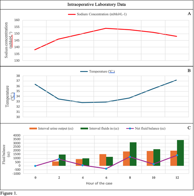
Figure 1
Given the intraoperative course, the anesthesiology team was concerned that the patient had DI, and 8 ug desmopressin was administered intravenously in the operating room, ten hours into the case. During surgical closure, ten and a half hours into the case, aggressive fluid resuscitation continued as delineated in figure 1C. Re-warming also continued with the same forced-air warmers, and room temperature was maintained at 23°C. The patient reached a core temperature of 35.5°C eleven hours after the start of the case and was successfully extubated. Urine output continued to remain high in the immediate post-extubation period. Her body temperature increased at an average rate of 0.55°C from hour 4 through 12 of the case. Figure 2 illustrates the patient’s temperature curve and highlights significant intraoperative management and events.
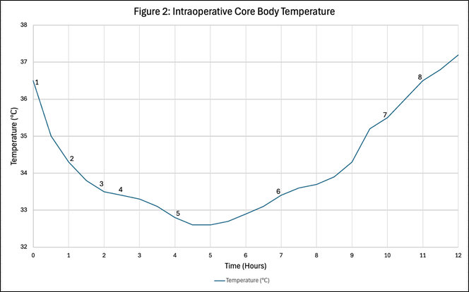
Figure 2 Core body temperature is charted for the 12-hour procedure with significant events as noted below:
Given the stark rise in sodium concentration and concerns for DI, the patient was admitted to the intensive care unit (ICU) for closer observation. Two hours after ICU admission, additional laboratory evaluation revealed a serum osmolality of 300 mOsm·kg-1, urine osmolality of 326 mOsm·kg-1, and a specific gravity of 1.006. In the ICU, the patient was given an additional 1 ug of desmopressin seven hours after the initial dose. A basic metabolic panel on postoperative day one (POD 1) revealed a sodium concentration of 141 mMol·L-1 and a urine osmolality of 634 mOsm·kg-1. The ICU team did not order a repeat serum osmolality. The average urine output decreased to 75 cc·hr-1 by POD 1. Following an uneventful recovery, the patient was discharged home POD 2 and instructed to follow up with endocrinology. The patient was lost to follow-up.
The body regulates its core temperature through a combination of autonomic and behavioral responses such as redirecting cutaneous blood flow, shivering, and sweating, depending on the environmental temperature. The hypothalamus controls this complex regulation.3 The hypothalamus normally maintains a core body temperature of around 36° - 37°C and triggers corrective responses outside of a temperature range known as the interthreshold range. GA widens the interthreshold range and disrupts homeostasis.7 Consequently, the body reaches abnormal temperatures without a compensatory response as central and peripheral control mechanisms are depressed.
The induction of GA precipitates profound peripheral vasodilation, leading to transdermal heat loss. This is the predominant mechanism of heat loss during the first hour of GA.7 The key to treating intraoperative hypothermia is prevention. This can be achieved with preoperative warming with forced-air warmers, which is efficacious in reducing both the likelihood and severity of intraoperative hypothermia.3,8 Forced-air warming is the most efficient method of rewarming after the patient becomes hypothermic.7
Active invasive options represent extreme but effective methods for re-warming. While not applicable to this case, cold bladder irrigation increases the risk of perioperative hypothermia in patients undergoing urologic surgery. One meta-analysis demonstrated that warm bladder irrigation reduced the occurrence of perioperative hypothermia in patients undergoing urologic procedures and represents a possible rewarming option for urologic procedures.9 Similarly, multiple case reports describe the utility of peritoneal lavage, particularly in extreme cases of hypothermia.10,11 Patients already had peritoneal access via a peritoneal dialysis catheter in these cases. These two routes of invasive rewarming may not be practical for all patients and surgical procedures, as was the case presented here. If peritoneal or bladder lavage is clinically appropriate, it offers a potential solution to refractory hypothermia in extreme cases.
There are a myriad of physiologic complications seen with hypothermia. Hypothermia alters pharmacokinetics, resulting in slower drug metabolism, consequently increasing the risk of medication overdose. Hypothermia alters thromboelastography, indicating dysfunction in clot formation, which increases the need for blood product transfusion.12,13
The differential diagnosis of polyuria includes medications, the diuretic phase of acute tubular necrosis, diabetes mellitus, DI, and primary polydipsia. Given the patient’s medical history, laboratory results, and acuity of polyuria, DI was suspected, and desmopressin was administered. The patient’s polyuria resolved following desmopressin and rewarming, supporting the diagnosis. With a rapid resolution of the patient’s polyuria, central DI was the likely diagnosis.
This case of DI describes a rare effect of hypothermia. ADH appears to be critical in regulating cold-induced diuresis; however, the underlying mechanism remains unclear. Animal studies suggest cold-induced diuresis is linked to reduced ADH activity, but it remains unclear whether this reflects lower ADH concentrations or renal resistance.5,14 No recent studies clarify the exact mechanisms by which this phenomenon occurs.
None.
None.

©2025 Madhani, et al. This is an open access article distributed under the terms of the, which permits unrestricted use, distribution, and build upon your work non-commercially.
