Journal of
eISSN: 2373-6437


Case Series Volume 17 Issue 5
1Department of Anesthesiology and Perioperative Medicine, UPMC Children's Hospital of Pittsburgh, USA
2Department of Surgery, UPMC Children's Hospital of Pittsburgh, USA
Correspondence: Mihaela Visoiu, MD, Department of Anesthesiology and Perioperative Medicine, UPMC Children's Hospital of Pittsburgh, 4401 Penn Avenue, Pittsburgh, PA, 15224, Pennsylvania, USA, Tel 412-692-5260
Received: September 01, 2025 | Published: September 23, 2025
Citation: Visoiu M, Joseph CA, Waltz P, et al. Subcutaneous hematomas following epidural catheter removal: two case reports. J Anesth Crit Care Open Acces. 2025;17(4):125-127. DOI: 10.15406/jaccoa.2025.17.00633
Epidural catheters are commonly used after surgery to manage pain effectively, but they carry risks, including complications like epidural hematomas and abscesses. Although these complications are rare, if left untreated, they can cause severe neurological damage. In contrast to epidural hematomas, intramuscular and subcutaneous hematomas are less risky but can be challenging to diagnose. This report discusses two cases of subcutaneous hematomas that occurred 24 hours following the removal of epidural catheters, highlighting the importance of close monitoring and collaboration among medical teams. Case 1 involves a young male with a complex medical history who underwent an exploratory laparotomy, while Case 2 involves a pediatric patient who underwent nephroureterectomy. The placement of the epidural catheters was uneventful. None of these patients had a coagulation disorder or received anticoagulation treatment. Both cases illustrate the diagnosis and management of subcutaneous hematomas, emphasizing the importance of early identification, differentiation from abscesses, and appropriate treatment. This report aims to increase awareness among healthcare providers about this uncommon but significant postoperative complication and to emphasize the importance of proactive management and patient education in ensuring a successful recovery.
Keywords: Epidural Catheter, Subcutaneous Hematoma, Bleeding Complications, Ultrasound, Back Swelling, Back Discoloration
Epidural catheters are often used to ensure adequate pain relief after major abdominal or thoracic surgeries. Despite their well-documented benefits,1 these catheters are not without risks. The high vascularity of the epidural space makes patients susceptible to bleeding complications, such as an epidural hematoma.2 An epidural abscess is another potentially serious, rare event.3-5 Both complications, if left untreated, can lead to spinal cord compression and permanent nerve damage.2,3
Intramuscular and subcutaneous hematomas are rarely encountered. While they pose no risk of neurological complications, distinguishing them from abscesses can be difficult. Additionally, evaluating their extension into the epidural space presents a challenge. These complications can lead to significant anxiety for patients, their families, and medical providers, as well as increased healthcare costs. This report presents two cases of subcutaneous hematoma following the removal of epidural catheters, highlighting the importance of close follow-up, interdisciplinary evaluation, and communication in the postoperative period. This article adheres to the guidelines laid by the Enhancing the Quality and Transparency of Health Research (EQUATOR) network. Written permission was obtained from the patients’ guardians to publish these case reports.
Case 1
A 22-year-old, 40.2-kilogram male, BMI 20.22, ASA 3, with past medical history of developmental delay, liver and small bowel transplant, immunosuppression, intra-abdominal abscess, diffuse colitis, was scheduled for exploratory laparotomy, proctocolectomy, and revision of the gastric tube. Preoperatively, the patient was hemodynamically stable, with normal liver function and coagulation studies.
To manage postoperative pain, a thoracic epidural catheter was inserted at the T11–T12 vertebral level using a loss-of-resistance technique. No complications were encountered during the placement, which required two attempts. Postoperatively, the catheter was infused with ropivacaine 0.2% at 6 mL/hour, and intermittent boluses of 4 mL every 4 hours. The pain was adequately controlled. The patient did not become coagulopathic or receive anticoagulant therapy. The epidural catheter was removed without any difficulties on the fourth postoperative day. No changes were noticed at the insertion site after epidural catheter removal.
About 24 hours after the epidural removal, the patient's family noticed a hard, non-erythematous, ovoid shape about 3 cm in width, localized at the epidural catheter insertion site (Figure 1A). The mass was tender to the touch, though it was unclear if the patient was experiencing back pain. The patient was afebrile, and neurologic examination was normal.
A bedside ultrasound revealed a well-circumscribed, ovoid collection of fluid and air in the subcutaneous fat, measuring 3.2 ×0.8 × 3.7 (caudal cranial x anterior posterior x transverse) cm in size (Figure 1B). There was a surrounding thickening of the subcutaneous fat without hyperemia. The sterility of the fluid collection could not be determined, and the ultrasound could not rule out the possibility of a deep fluid collection. Concerns were raised about a localized infection or hematoma. On the third day, the mass seemed softer, and later an area of redness and discoloration developed around the prior needle insertion site (Figure 1A).
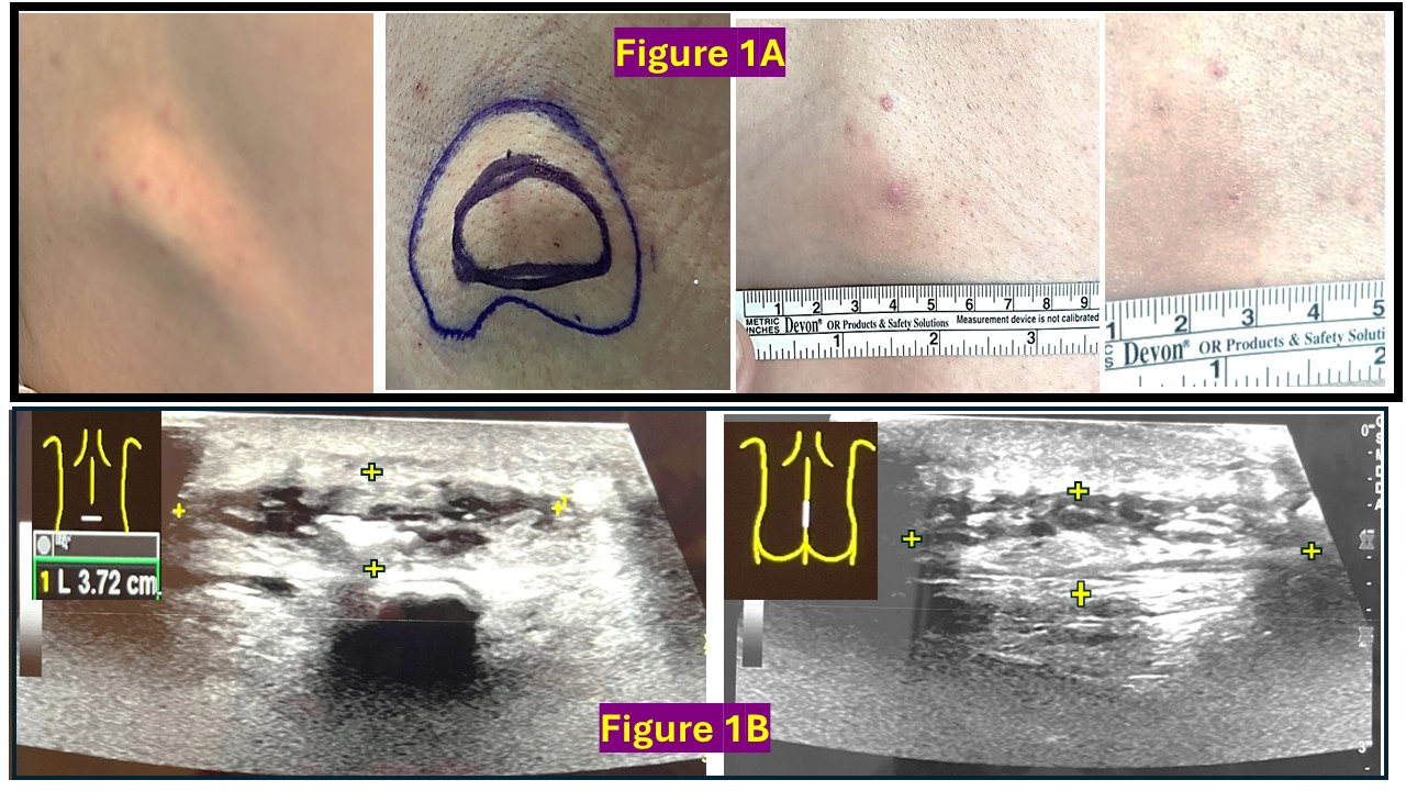
Figure 1A Pictures of the hematoma of the first case, from the first day when it was noticed until almost complete resolution.
Figure 1B Ultrasound image of the hematoma of the first case, with the probe placed in both transverse and sagittal orientations.
The absence of any inflammatory signs made the possibility of a localized or epidural abscess less likely. A local biopsy of the mass was considered; however, this procedure would require additional sedation and was not desirable. In collaboration with the primary team and the interventional radiology physician, the pain physician decided to closely monitor the patient's back mass, focusing on the local physical exam, pain behavior, vital signs, and inflammatory markers. The family was informed that the diagnosis was a localized subcutaneous hematoma and was instructed to apply warm compresses and apply topical diclofenac gel. The patient was discharged a week later, with the mass gradually resolving. Telephone follow-ups with the mother confirmed a gradual reduction in swelling, with complete resolution confirmed 15 days after the initial presentation.
Case 2
A 5-year-old girl, weighing 18.4 kg, with a BMI of 14.96, ASA 3, and a history of left Wilms tumor, was scheduled for left nephroureterectomy, lymph node sampling, and port placement. Preoperative assessment included a normal coagulation profile and a platelet count exceeding 150,000/µL.
For postoperative pain control, a thoracic epidural catheter was inserted at the T11-T12 vertebral level using the loss-of-resistance technique, which required one attempt. No complications were encountered during the placement. Postoperatively, the catheter was infused with ropivacaine 0.2% and clonidine 1 mcg/ml at 4 ml/hour and intermittent boluses of 2 ml every 4 hours. The pain was adequately controlled. Daily inspection of the epidural insertion site revealed no skin changes, tenderness, or discharge. The patient did not become coagulopathic or receive anticoagulant therapy during the hospital stay. The epidural catheter was removed on postoperative day two, one day before the patient’s discharge.
Twenty-four hours after discharge, the child's mother noticed swelling and redness at the epidural site, along with a small black dot. Over the next day, the affected area increased to about 2.5 cm in diameter, with a black scab forming in the center surrounded by erythema (Figure 2A). Although the child had no fever or neurological deficits, the mass was tender to the touch, raising concerns about a localized infection. The parent was instructed to bring the child to the emergency room.
On arrival, the physical exam revealed a red, tender, and firm back mass. There were also ecchymotic skin changes, with no discharge. Ultrasound revealed a large, complex collection within the soft tissues of the midline of the spine, with associated fat stranding and gas locules. The collection measured 5.6 x 0.7 x 3.2 cm (caudal cranial x anterior posterior x transverse) (Figure 2B). There was hyperemia of the adjacent tissues.
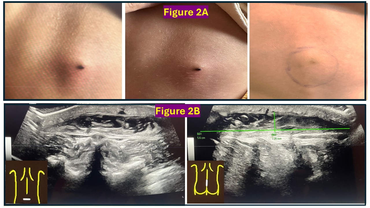
Figure 2A Pictures of the hematoma of the second case, from the first day when it was noticed until almost complete resolution.
Figure 2B Ultrasound image of the hematoma of the second case, with the probe placed in both transverse and sagittal orientations.
No definite tract was visualized to extend from the collection into the spinal canal. Concerns were raised about a hematoma or an abscess. The absence of any inflammatory signs made the possibility of a localized or epidural abscess less likely. A spine MRI and a needle aspiration of the mass’s content were considered; however, both procedures would require sedation and increase the hospital stay and medical expenses. In collaboration with the surgeon, oncologist, and emergency medical team, the pain physician decided to monitor the patient's mass and discharge her home with close follow-up. The family was informed that the diagnosis was a localized subcutaneous hematoma and was instructed to apply warm and cold compresses and topical antibiotic cream. A few days later, the back scab fell off, followed by a bloody discharge, with a significant decrease in mass size (Figure 2A). The parents reported a complete resolution of the swelling 10 days after it was discovered.
Subcutaneous hematomas following the removal of epidural catheters are rare and have not been previously reported as a complication. However, hematomas at block sites have been documented after continuous erector spinae plane blocks,6,7 single injections of quadratus lumborum blocks,8 and continuous paravertebral nerve blocks.3 Williamson et al. described a local hematoma that was noticed upon removing an erector spinae plane catheter and continued to grow over subsequent days.6 The patient had normal coagulation levels and received postoperative subcutaneous heparin. Joseph et al. reported a large hematoma that possibly developed two days after removing an ESP catheter.7 This patient received anticoagulation therapy both during catheter maintenance and after its removal, and became coagulopathic after being discharged home.
In our cases, no immediate changes were noted upon catheter removal, but hard, tender masses developed insidiously over the next 24 hours. Neither patient had underlying coagulopathies or received anticoagulation during their hospital stay. The exact cause remains unclear, but it is believed that vessel injury during catheter placement led to subsequent bleeding post-catheter removal, possibly due to the catheter obstructing the initial needle punctures.
In both cases, the primary challenge was differentiating hematomas from localized abscesses and ruling out extension into the epidural space. While needle aspiration or incision and drainage could have been considered to assess fluid sterility, the absence of inflammatory signs made a localized infection less likely. The presence of bruising around the needle insertion site, along with bloody discharge and subsequent mass reduction, made a hematoma diagnosis more probable.
Walker et al. described the potential extension of the blood collections into the epidural space. They reported swelling at paravertebral catheter sites, with MRI confirmed hematoma extension.3 However, our patients showed normal neurological exams, reducing concern for extension that would necessitate additional investigations.
After performing ultrasounds, we discussed our concerns and potential investigations with the patients' caregivers and the surgical and primary care teams. We provided reassurance and followed up with the patients' parents and medical providers to ensure they were informed and comfortable with the plan of care.
These case reports underscore the importance of discussing potential complications, such as subcutaneous hematomas after epidural catheter removal. Practitioners should remain available for questions and follow-up after these catheters are removed.
In conclusion, while subcutaneous hematomas after epidural catheter removal are rare, they should be considered as a possible complication. Early identification, appropriate management, close follow-up, and family education are key to preventing unnecessary interventions and ensuring favorable outcomes.
No competing interests or funding declared.
None.
None.

©2025 Visoiu, et al. This is an open access article distributed under the terms of the, which permits unrestricted use, distribution, and build upon your work non-commercially.
