Journal of
eISSN: 2574-9943


Case Report Volume 7 Issue 4
Maxillofacial Surgery Department La Paz Hospital Madrid, Spain
Correspondence: Carolina Cuesta Urquia, Hospital Universitario La Paz, Paseo de la Castellana 261, 28046 Madrid, Spain, Tel +34697740997
Received: December 05, 2023 | Published: December 26, 2023
Citation: Cuesta Urquia C, Moreiras Sánchez AD, Pampín Martínez MM, et al. Surface scanning and 3D printing for optimized partial auricular reconstruction. J Dermat Cosmetol. 2023;7(4):154-156. DOI: 10.15406/jdc.2023.07.00253
We present the resolution of a case of a 50-year-old patient who presented for excision and reconstruction of a malignant skin lesion at the auricular tubercle of the right helix. The patient reported great concern for the aesthetic outcome. The auricular helix has a unique projection, relief, consistency, and size determined by the contralateral side. Its reconstruction is a surgical challenge and requires sophisticated techniques to achieve good results. Therefore, in cases where we are looking for precision and predictability, 3D technology is a great help for the surgeon as it allows pre-surgical patient-specific planning and the printing of sterilisable models that serve as surgical 3D guides. This case was solved with a two-stage reconstruction. First, the lesion was excised, and a retro auricular advancement flap was placed. In the second stage, the placement of cartilage was sectioned using a customised surgical model obtained from the scanning of the contralateral ear surface. This way, the cartilage donor area was optimised, choosing the most similar shape and size with great precision.
Keywords: partial ear reconstruction, surface scan, 3D printing, surgical planning
In the facial region, any increase in the excised skin surface increases the complexity and morbidity of reconstruction. These characteristics are even more marked in the auricle, particularly the helix, due to its unique anatomy and relief, decreasing reconstructive possibilities. The main indication for the post-auricular advancement flap is the reconstruction of helix auricular defects. In this case, oncological resection required excision of the cartilage (Figure 1); reconstruction to preserve the consistency of the helix required cartilage graft. Therefore, 3D technology assisted the surgeon with precise and optimised cartilage harvest to acquire precision and predictability.
Main body
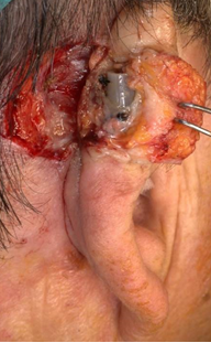
A 50-year-old patient presented with a malignant skin lesion at the auricular tubercle of the right helix. The patient reported great concern for the aesthetic outcome. Oncological resection margins required excision of the cartilage (Figure 1). The case was solved with a two-stage reconstruction. First, the lesion was excised, and a retro auricular advancement flap from postauricular skin was attached to the lateral margin of the auricular defect. To preserve the relief and prevent collapse, a silastic tube was inserted under the skin flap fixed with silk thread (Figure 2).

Figure 2 Second stage surgery: removal of silastic tube previously inserted under the skin flap fixed with silk thread.
In the second stage, 3D technology was used to choose the most accurate donor site for cartilage graft. Patient-specific model creation is already a routine in our department. The protocol detailed by Rodriguez-Arias et al.,1 is adapted to patient needs. It consists of four stages: surface scanning (Figure 3), model design and preparation, 3D printing, and surgery (Figure 4).
The first step was the Surface Scanning of auricles, which consisted of the image acquisition of the healthy and operated ear of the subject. A structured-light surface 3D scanner allowed the capture of the detailed features of the auricular structure (Artec Eva, Artec Group, Luxembourg) with a 3D resolution of up to 0.2 mm at a 16 fps reconstruction rate. The obtained point cloud data were processed in the native Artec Studio 16 Professional software (Artec Group, Luxembourg).
This data was converted to two different meshes imported as STL files and used on the software Meshmixer (v 3.5, Autodesk Inc., San Rafael, CA, USA). The mesh of the operated ear indicated the defect's size, and the healthy ear's mirrored mesh was used to create a digital model of the cartilage graft.
Finally, the 3D model representing the cartilage graft to be harvested in actual size and curvature (Figure 4) was printed in Surgical Guide resin before surgery. This model was sterilized, and during the second stage surgery, was placed in different parts of the auricle, finding the triangular fossa to be the most likely to the recipient site. This way, the cartilage donor area was optimized, choosing the most similar shape and size with great precision.
The reconstruction preserved the consistency and shape of the helix (Figure 5) with minimal repercussions on the donor site.
Three-dimensional (3D) planning and personalized surgery have emerged as revolutionary approaches in the field of medical interventions, particularly in complex surgical procedures. The utilization of advanced imaging technologies allows for the creation of detailed and patient-specific 3D models, enabling surgeons to meticulously plan procedures prior to the actual surgery. This tailored approach enhances precision and accuracy, minimizing the risk of complications and optimizing outcomes. This case could have been resolved without this technology, but the personalized surgical guide, fabricated based on the patient's anatomy, contributes to improved surgical efficiency and reduced operative times. Furthermore, the successful implementation of 3D planning and personalized surgery is time-consuming and requires practice. Once a multidisciplinary collaboration involving advanced technological infrastructure is available, there is no denying the benefits of technology. Despite these challenges, the continuous development and integration of these techniques hold great promise in advancing the field of surgical practice.2
While acknowledging that the case could have been managed without 3D technology, the personalized surgical guide proved instrumental in enhancing surgical efficiency and reducing operative times. The preservation of the helix's consistency and shape with minimal repercussions on the donor site underscores the potential of 3D planning and personalized surgery in overcoming the intricacies of facial reconstruction.
Despite the challenges associated with time consumption and the need for multidisciplinary collaboration and advanced technological infrastructure, the benefits of 3D planning and personalized surgery in complex cases like this one are undeniable. As technology continues to evolve, its integration into surgical practice holds great promise, offering a pathway to further advancements in the field of facial reconstruction and beyond.
None.
The authors declare there is no conflicts interest.

©2023 Cuesta, et al. This is an open access article distributed under the terms of the, which permits unrestricted use, distribution, and build upon your work non-commercially.