Journal of
eISSN: 2373-4345


Case Report Volume 15 Issue 1
1DDS, PhD, Responsible for Research, Istituto Stomatologico Toscano, Italy
2Prof, Department of Stomatology, Faculty of Dentistry, University of Seville, Spain
3DDS, Oral Surgery Resident, Università Vita Salute San Raffaele, Italy
4Prof and Director at Istituto Stomatologico Toscano, Italy
Correspondence: Enrica Giammarinaro, DDS, Oral Surgery Resident, Università Vita Salute San Raffaele, Italy
Received: January 22, 2024 | Published: February 13, 2024
Citation: Marconcini S, Velasco-Ortega E, Giammarinaro E, et al. Immediate implant in the esthetic area when immediate loading is not an option: a clinical case report. J Dent Health Oral Disord Ther. 2024;15(1):18-21. DOI: 10.15406/jdhodt.2024.15.00610
Aims: Tooth extraction in the frontal area is associated with significant bone resorption, especially at sockets presenting with a thin buccal wall, such as most of the maxillary incisors. In the case of favorable residual hard and soft tissue anatomy, immediate implant placement in the esthetic area might be an opportunity to drive the alveolus healing course in the desired direction.
Case presentation: In this clinical case, a fractured upper central incisor was replaced by an immediate two-piece implant covered with a healing screw. After 3 months, the implant was restored. The facial and interdental soft tissue was maintained with appreciable success at the 3-year follow-up visit.
Conclusion: When immediate loading of the implant is not an option and the fresh extraction socket presents with fair soft tissue quality, the use of an interim healing screw might help sustain and prevent the shrinkage of peri-implant tissues. This simple technique might be a valuable option when there is no indication for adjunctive hard or soft tissue grafting or immediate loading feasibility conditions.
Keywords: connective tissue biology, implantology, wound healing, ridge preservation, fibroblast(s), cosmetic dentistry
Immediate implant placement in the anterior maxilla is challenging because of the esthetic expectation from both the patient and the clinician.
Tooth extraction leads to sensible bone remodeling:1,2 severe alveolar bone remodeling affects the entire socket in both the vertical and horizontal directions, with volumetric shrinkage whose centroid is shifted lingually3 Most authors attributed this change to the loss of periodontal ligament; however, bone remodeling might depend on the residual anatomy (tooth site, tooth axis and inclination, bone plate thickness) more than on the disruption of bundle bone.5 The preservation of ridge topography might be more important than the integrity of the socket.4 Different techniques have been proposed to improve the residual anatomy or to prevent hard and soft tissue remodeling: flapless intervention, proper implants axis orientation, bone compacting/dislocation, soft tissue augmentation, and favorable abutment geometry.6–8
This report describes a simple protocol for the management of the peri-implant mucosa soon after implant insertion at post extraction sites whenever immediate loading is not an option.
Clinical presentation
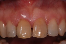
A woman aged 40 years presented to the Tuscan Stomatologic Institute (Forte dei Marmi, Italy) with pain caused by a fracture of the maxillary right central incisor. The patient provided informed written consent before treatment. A thorough intraoral examination, study casts, a radiographic evaluation, and photographic documentation were performed (Figures 1, 2). The 3D scans revealed sufficient apical bone height that would have allowed implant primary stability.

Figure 1 BEFORE: Initial Presentation. Clinical appearance of central incisors with a low fistula at the element 1.1 and overall unsatisfactory esthetics of the frontal group with evident dyscromia.
Based on clinical examination and diagnostic tools, a few treatment plans were proposed and fully explained to the patient. The extraction of element 1.1 and the placement of an immediate implant were chosen among other rehabilitation options. Analog impressions of the maxilla and mandible and registration of the bite were taken. The esthetic of the contralateral element was restored as well (element 2.1).
Case management
On the day of the surgery, amoxicillin+clavulanic acid (2 g) was started two hours before the intervention. Under local anesthesia, without raising a flap, the surgeon (SM) removed the tooth carefully and performed meticulous cleaning and socket debridement.
After careful probing of the socket, the surgeon drilled through the new implant bed in a slightly palatal position and without raising a flap to avoid impairment of the periosteal blood supply. The implant (length 11.5, diameter 3.80, Kohno, Sweden & Martina) was seated at an iuxta-crestal position, and the buccal gap was filled with a collagen sponge (Gingistat, Vebas).
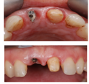
It was decided to avoid immediate loading, but the implant was left unsubmerged by means of a healing screw (Figure 3). The screw supported the soft tissue at the subcritical contour. The critical contour was sustained with the polished cervical portion of the tooth that was splinted to the adjacent teeth as a provisional restoration. The patient was given instructions about personal oral hygiene and postoperative behavior. The healing period was uneventful. Three months after surgery, a screwed definitive restoration was delivered for the immediate implant.
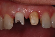
The healing of the surrounding tissues is shown after 3 months (Figure 4). At this time, soft tissue appeared pink and thick with an abundant profile, optimal for conditioning via provisional restoration. For the final restoration, a precision impression was taken with a double silicon material. A custom zirconia abutment was delivered (Figure 5). At the 3-year follow-up visit, the radiograph showed stability of the marginal bone (Figure 6). Clinically, the implant showed optimal esthetics and stability of the surrounding tissues with overall mimesis and color integration (Figure 7).

Figure 4 The healing of the surrounding tissues is shown after 3 months with satisfactory appearance, bulky anatomy, and conditioned tissue.

Figure 5 Detail of the custom zirconia healing abutment delivery and contralateral tooth preparation.
In the present clinical case, a healing screw and the splinted crown of the fractured tooth were used to sustain soft tissues immediately after implant placement. It must be noted that the extractive alveolus had a favorable residual anatomy with a complete 4-walled structure.
Several techniques have been proposed to preserve the anatomy after tooth extraction, and to do so by means of immediate implant treatment is one of the most challenging choices.9 Post extractive implant placement requires an expert operator, proper tridimensional implant positioning, careful management of the residual buccal bone, and the choice of the right time-lapse between implant placement and restoration.
The extractive alveolus is a composite wound that includes different cell types. The migration of fibroblasts through the extracellular matrix during the initial phase of healing is a fundamental component of wound contraction and remodeling.10 In particular, during the first 15 days; the coronal portion of the socket is exposed to significant centripetal contraction.
Most authors have recommended the use of bone grafts to fill the gap between the socket walls and the implant.11 However, where there are no large bone defects; the implant bed is a space-keeping bone defect and, therefore, a spontaneous-healing one. Thus, the main actor in the repair process would be a stable blood clot.
It has been reported that to maintain the stability of the facial soft tissue level, the buccal wall should be at least 2 mm thick.12 Unfortunately, a scenario with more than 2 mm thickness is rare, and facial bone loss and ridge deformation are common in the esthetic area. Therefore, it is highly recommended to improve the soft tissue design as early as possible. Multiple surgeries at the same site might lead to greater bone loss, soft tissue recession, and rigid-non moldable peri-implant mucosa.
The immediate restoration of post extraction implants may provide greater support to the facial and interproximal soft tissues in the healing period, leading to improved esthetic outcomes. The systematic review by Qi Yan and colleagues suggested that post extraction implants with immediate restoration in the esthetic area result in similar outcomes compared with conventional loading protocols.13 These results have been recently confirmed by Arora and Ivanovski, who compared immediate restoration and conventional restoration protocols for immediately placed implants in the anterior maxilla over a mean 3-year follow-up period.14
The results of a recent study suggested that the use of connective tissue grafts is not mandatory to achieve pleasant aesthetic outcomes in the case of immediate implant placement with immediate nonfunctional restoration.15
Similar to immediate restoration, the healing screw or abutment would act as a geometrical stop for the soft tissue. Of course, the same result could be achieved with a customized abutment and/or immediate loading procedure.
The present case report described a single patient with favorable residual anatomy and few local risk factors. Immediate nonfunctional restoration of post extraction implants should always be considered the first choice for preventing tissue remodeling.
This clinical case report suggests that immediate implant placement is an esthetic choice, especially if mechanical support is provided for marginal peri-implant hard and soft tissues and, in selected cases that may be achieved with a simple healing screw. Due to the nature of the present study, further research based on a larger population and with a more extended follow up will be necessary will be necessary in order to validate this technique
SM, EVO, EG, UC: 1) substantial contributions to conception and design, or acquisition, analysis, or interpretation of data; 2) drafting the article or revising it critically for important intellectual content; 3) final approval of the version to be published; and 4) agreement to be accountable for all aspects of the work in ensuring that questions related to the accuracy or integrity of any part of the work are appropriately investigated and resolved.
This research received no external funding.
Informed consent was obtained from the subject involved in the study.
None.
The authors declare that there are no conflicts of interest.

©2024 Marconcini, et al. This is an open access article distributed under the terms of the, which permits unrestricted use, distribution, and build upon your work non-commercially.