Journal of
eISSN: 2373-4426


Case Report Volume 14 Issue 1
1Guelmim Regional Hospital Center, Morocco
2Department of Urology A, Ibn Sina University Hospital Center, Morocco
Correspondence: Youssef Kadouri, Department of Urology, Guelmim Regional Hospital Center, Guelmim, Morocco
Received: January 09, 2024 | Published: January 23, 2024
Citation: Kadouri Y, Lakssir J, Sahnoun Z, et al. Surgical repair of a ventral penile fracture: a case report. J Pediatr Neonatal Care. 2024;14(1):30-31. DOI: 10.15406/jpnc.2024.14.00537
Ventral fracture of the penis, although rare, is a medical emergency requiring immediate attention. This injury usually results from direct trauma to the erect penis and can lead to serious complications if not treated promptly. Initially, it requires an attentive clinical assessment and habitually, it is completed with ultrasound to characterize more the fracture and help with the surgical approach. Through this case report, we will present a case about a delicate localization of the fracture, and we will discuss the causes, symptoms, diagnosis and treatment options for this condition.
Ventral fracture of the penis is defined by a rupture of the tunica albuginea, a fibrous envelope surrounding the corpora cavernosa of the penis causing a blood flow and hematoma formation.1 Urethral lesions are associated in 10 % of cases. This type of injury usually occurs during sexual intercourse, or less frequently secondary to a fall or direct trauma to the erect penis causing a sudden increase in pressure within the cavernosal.
Although relatively rare, this injury requires urgent medical attention, and an immediate surgical repair considered the gold standard2 due to the risk of long-term complications.
A 43-year-old patient with no medical history, married, and father of two children, who presents to the emergency room for the sudden appearance of a lump at the base of the penis. The questioning reveals a cracking sound occurring during sexual intercourse with his partner, raising suspicion for the diagnosis. Clinical examination reveals a painful formation at the base of the penis with an absence of the typical eggplant-like appearance associated with the diagnosis (Figure 1).
A supplementary ultrasound confirms the diagnosis of penile fracture by revealing the presence of a fracture line in the tunica albuginea of the corpus cavernosum, along with a hematoma measuring 3 cm in the corresponding area.
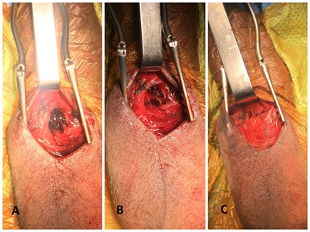
The patient was admitted to the operating room within 24 hours. An elective incision allowed direct access to the fracture at the base of the penis on its ventral side (Figure 2A). Surgical exploration, following hematoma evacuation (Figure 2B), revealed a transversal fracture of 20 mm repaired using Vicryl 3-0 while the structure of the anterior urethra was intact (Figure 2C).

Figure 2 Image showing the intraoperative appearance of a penile fracture,
A: Elective approach to the fracture line.
B: Appearance after hematoma evacuation.
C: Transversely repaired fracture line with Vicryl 3.0.
The immediate postoperative course was simple, with the patient discharged on the second day with pain relievers and anti-androgens to prevent the occurrence of erections that could lead to suture dehiscence. Long-term follow-up at 1 and 6 months showed no evidence of induration or fibrosis at the incision site, and erectile function was preserved.
Penile fracture is an uncommon situation, representing a rare circumstance for emergency department that might be undiagnosed, as it can lead to potentially significant functional complications. Its incidence is rare, with occurrences for example in North America ranging from 0.29 to 1.36 per 100,000 people.3
The occurrence of the fracture line is often observed on the right side of the corpus cavernosum on the proximal shaft or midshaft, accounting for 72%, and the addition of urethral injury is found in 10% of cases. However, the occurrence of a fracture line at the base of the penis, as in our case, is 31%.3
The occurrence of penile fracture is associated with direct trauma occurring during an erection. This may happen during vigorous sexual intercourse, abrupt movements, or forceful impacts against the erect penis, leading to a rupture of the tunica albuginea.4 This rupture causes blood to leak from the corpora cavernosa, resulting in severe pain and hematoma. In the United States, the most common cause is typically related to sexual activity. However, in the Middle East, it is often attributed to a penile manipulation known as "Taqaadan," performed with the intention of inducing detumescence forcibly.3
The clinical diagnosis requires careful attention, relying on a thorough clinical examination combined with the patient's history. It is noteworthy that the reported symptoms by the patient are highly characteristic, including hearing a popping or cracking sound associated with pain, rapid detumescence, swelling, and penile deformity.5 Additionally, a skilled examination may allow the identification of the fracture location through a 'rolling sign,' detecting clots within the torn tunica. This corresponds to the most tender point of the shaft, aiding in the surgical approach.3
In uncertain situations, imaging can provide significant assistance in diagnosis. First and foremost is ultrasound, considered a rapid, simple, and cost-effective examination, allowing the identification of fractures in 86% of cases.6,7 However, it has some limitations, particularly in the presence of a significant hematoma that may obscure the fracture line, and its efficacy relies on the experience of the radiologist. As for MRI, it offers high sensitivity with a detection rate of fracture between 98 and 100%1,7 but remains a costly and generally inaccessible examination, especially in emergency situations.
Other complementary tests may be indicated, such as retrograde urethrography when there is suspicion of urethral involvement. It is possible to visualize the urethra through ultrasound by a retrograde technique where a saline solution or a lubricant anesthetic jelly is introduced into the urethra using either a Foley catheter or a syringe directly placed in the meatus.8 This should be considered in the presence of a clot in the urethral meatus, urethral bleeding, or urinary retention.3
However, imaging should not delay therapeutic intervention. In many cases, imaging may be entirely unnecessary when the clinical presentation is evident. Surgical treatment is the gold standard, involving hematoma evacuation to reduce the duration of local inflammation and repair of the tunica albuginea defect. Conservative medical treatment is to be avoided, as it is associated with a higher incidence of late-onset morbidity, such as Peyronie's disease.3,4
At the end, if not treated promptly, penile fracture can lead to serious complications, including erectile dysfunction, abnormal penile curvature and psychological problems related to sexual function. However, with appropriate surgical intervention, most patients can regain normal erectile function and minimize sequelae.
Penile fracture requires a careful examination, and in some cases, imaging may improve surgical management and counselling by confirming the diagnosis, locating and evaluating the extension of the injury. Urethral involvement should be considered and investigated based on its indicative signs. Timely identification and swift surgical intervention are pivotal for achieving favorable outcomes. Conversely, delayed presentation may pose various challenges.
None.
Our institution does not require ethical approval for reporting individual cases or case series.
All the authors have contributed to the production of this book by studying the medical records, and ensuring the interventions, the care discussions as well as the reading of the book after its writing.
Written informed consent was obtained from the patient(s) for their anonymized information to be published in this article.
Consent was obtained from the patient to publish this case report and accompanying images.
Ethical approval is not applicable. The case report is not containing any personal information.
This study received no funding from any resource.
The authors declare that there are no conflicts of interest.

©2024 Kadouri, et al. This is an open access article distributed under the terms of the, which permits unrestricted use, distribution, and build upon your work non-commercially.