MOJ
eISSN: 2381-179X


Case Report Volume 13 Issue 2
1Orthopaedics Surgery Resident, Universidad Finis Terrae, Chile
2Pediatrics Orthopaedic Surgeon, Hospital Dra. Eloisa Díaz, Universidad de Santiago de Chile, Chile
3Pediatrics Orthopaedic Surgeon, Hospital Dra. Eloisa Díaz, Universidad de Santiago de Chile, Chile
Correspondence: José Torrealba Araujo, MD, Universidad Finis Terrae, Providencia, Santiago, Chile
Received: June 07, 2023 | Published: June 19, 2023
Citation: José TA, Lautaro CT, Francisco CZ. Use of the 3D printing in surgical planning of the femoroacetabular impigement of the right hip. MOJ Clin Med Case Rep. 2023;13(2):51-53. DOI: 10.15406/mojcr.2023.13.00436
Femoroacetabular impingement occurs when the femoral head has abnormal contact with the acetabulum of the pelvic bone. This condition predisposes the hip joint to instability, chondral or labral lesions and progressive osteoarthritis. Legg-Calvé-Perthes disease occurs frequently in children between 2 and 12 years of age. Three-dimensional printing technology is being used in all preoperative planning, allowing understanding of the lesions, measure the size of the CAM lesion and could improve the precision, operative settings and reduce the use of x- rays.
Keywords: three-dimensional printing, secular injuries, femoroacetabular impingement, surgical hip dislocation, Legg-Calvé-Perthes
In the recent past, a group of young people, with or without history of previous affection of the hip, complained of pain in the inguinal region, during or after physical activities or after long sitting periods. Paradoxically, the physical examination was poor and the radiographs were interpreted as a normal aspect or, in some cases, presented alterations consistent with sequelae from previous illness, such as Legg-Perthes or slipped capital femoral epiphysis, but that did not explain the symptoms in the light of the knowledge at the time. As a result, there was no specific diagnosis and therapy; the recommendation was symptomatic treatment and restriction of physical activities. However, in some cases, there was long term evolution to articular degeneration.1,2 Legg-Calve-Perthes disease (LCPD) is defined as remodeling of the proximal femoral epiphysis during growth after idiopathic ischemia. At stake is the onset of hip osteoarthritis, occurring more or less rapidly depending on the sphericity and congruency of the femoral head. Treatment of FAI in LCPD is specific. Simple resection of the anterolateral offset of the femoral head is relatively simple and only minimally invasive, and seems to give satisfactory results even if this needs to be confirmed.3 Three-dimensional printing (3D) is an additive manufacturing technique, which through digital models of a patient, obtained through different techniques such as computed tomography (CT), allows the manufacture of custom made and specific structures for each patient, in real size, by using plastic materials.4 Undoubtedly, a specialty of medicine in which it is generating great interest is Traumatology and Orthopedics, through the printing of orthoses/prostheses, surgical instruments and anatomical models.5
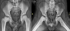
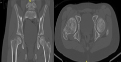
A 12-year-old male patient, who began medical controls in our service in 2018, diagnosed with Legg-Calvé-Perthes disease of the right hip (Figure 1), presenting pain when standing, agitated gait, and claudication. In 2022 in tomographic control (Figure 2) and CT with 3D reconstruction (Figure 3) that evidence right coxa magna and alteration of the femoral sphericity with CAM type morphology, associated with pain and limitation of range of motion, was the reason for which it was decided to perform surgical intervention. Physical exam: Pain and limitation of range of motion (right/left flexion 100°/100°, right/left internal rotation 28°/45°, right/left external rotation 12°/20°, limited abduction due to pain in the right lower extremity).

Figure 1 AP and Lowestein Pelvis X-ray from 2018, showing Legg-Calvé-Perthes disease of the right hip.

Figure 2 Computed Axial Tomography of the hip from the year 2022, showing evidence of the right coxa magna and alteration of the femoral sphericity of CAM type morphology.
Complementary exams
Diagnosis
Right femoroacetabular impingement with CAM-type morphology due to sequelae of Legg-Calvé-Perthes disease.
Treatment
Preoperative planning is carried out through imaging studies, digital segmentation and 3D printing of the right hip (Figure 4), where the CAM-type morphology lesion can be evidenced. In the programmed operative room, under general anesthesia, in left lateral decubitus, an anterior approach to the hip is performed and dissection by planes up to the fascia, which is excised. Subsequently, an osteotomy of the greater trochanter is performed, release of the gluteus medius, the joint capsule is released, observing the femoral head with healthy articular cartilage; capsulotomy is performed and the Impingement test is performed under direct vision. Then, controlled hip dislocation is performed using a flexion and adduction maneuver, and resection of the anterolateral portion of the coxa magna is performed with an osteotome and shaver (DePuy-Synthes). Finally, hip reduction is performed and compliance is visualized under fluoroscopic vision in two planes, the greater trochanter is transposed and fixed with 2 6.5 mm cannulated screws (DePuy-Synthes), reduction compliance is visualized with fluoroscopic vision, and it is verified with direct visualization with a 3D model, suture is performed and the post-surgical cleaning with sterile dressings and gauze is performed, ending the procedure without complications. Radiological control is performed (Figure 5) and due to satisfactory postoperative evolution, medical discharge is decided after 24 hours. Radiographic control is performed at 6 months, where appropriate bone healing is evidenced (Figure 6).
Femoroacetabular impingement syndrome is a clinical entity that has aroused interest in the last 15 years and has been related to the development of hip pain and early osteoarthritis.6 This mechanical condition predisposes the hip joint to dynamic instability, localized joint overload, impingement, or a combination of these characteristics, which become potential mechanisms for the development of chondral and labral lesions, and progressive osteoarthritis in most patients. The hips, thus altering the patient's physiological range of motion and interfering with their daily activities.7 Legg-Calvé-Perthes disease is a term reserved for idiopathic avascular necrosis of the juvenile hip, currently its etiology is somewhat uncertain, and it usually occurs more frequently in the pediatric population between 2 and 12 years of age.8 Currently, PCL disease is a term reserved for idiopathic avascular necrosis of the juvenile hip, which can be associated with different pathophysiological mechanisms regarding its etiology, which generates delays in diagnostic suspicion as well as in treatment.8
Three-dimensional printing is emerging as a promising technology to personalize anatomical models, allowing us to understand the anatomy of our patients in a much more concrete way than traditional radiological images. These models provide an accurate representation of the lesions, becoming in some cases an indispensable tool for preoperative planning.9 This is a growing technology, used today in the field of engineering, architecture, entertainment, education, in addition to its use in various areas of health, which has had an increase in the number of publications in indexed journals per year since 2013.10 Studies, such as that of Chen C et al.,11 have shown that by using this technology in preoperative planning, operating time, blood loss, and the use of X-rays are significantly reduced.
Three-dimensional printing is a technology that could be an excellent contribution in all the operative setting, planning, and the diagnosis, therefore it is very likely that we will continue to see an increase the work to generating publications in the coming years. In this particular case, the Three-dimensional model improves the direct visualization of the CAM lesion, and collaborate with the surgical team to realize the proper procedure.
None.
Authors declare that there is no conflict of interest.

©2023 José, et al. This is an open access article distributed under the terms of the, which permits unrestricted use, distribution, and build upon your work non-commercially.