eISSN: 2377-4304


Case Report Volume 13 Issue 3
1Resident doctor of the specialty of Gynecology and Obstetrics. Maternal and Perinatal Hospital ‘’Mónica Pretelini Sáenz’’ Toluca, Mexico
2Physician attached to the Maternal-Fetal Medicine Service. Maternal and Perinatal Hospital ‘’Mónica Pretelini Sáenz’’, Toluca, Mexico
Correspondence: Oscar Alberto Gómez Morales, Resident doctor of the specialty of Gynecology and Obstetrics. Maternal and Perinatal Hospital ‘’Mónica Pretelini Sáenz’’ Toluca, Mexico
Received: June 10, 2022 | Published: June 20, 2022
Citation: DOI: 10.15406/ogij.2022.13.00646
A clinical case of a 24-year-old female is presented, which by first-trimester obstetric ultrasound data suggestive of an incomplete mole is detected, first-trimester screening is performed, reporting a fetus without structural alterations, pregnancy interruption is offered, the same as the patient refuses, pregnancy is continued until the end of 37 weeks of gestation; where a fetus is obtained phenotypically without structural malformations.
Gestational trophoblastic disease is defined as a rare complication of pregnancy characterized by abnormal proliferation of trophoblastic tissue. Which includes a wide spectrum of clinicopathological entities ranging from benign GTD (Complete Hydatidiform Mole and Partial Hydatidiform Mole) to malignant pathologies (Invasive Hydatidiform Mole, Choriocarcinoma, Placental Site Tumor and Epithelial Trophoblastic Tumor) also known as Gestational Trophoblastic Neoplasia ( NTG).1
The incidence of gestational trophoblastic disease varies by geographic area. In Mexico it is 2.4 per 1000 pregnancies, the incidence of invasive mole occurs in 1 of every 40 molar pregnancies and 1 of every 150,000 normal pregnancies. GTD can occur after a molar pregnancy, a normal pregnancy, or an ectopic pregnancy.2
Female patient, 24 years old, primigravida, pregnant at 9.4 weeks of gestation, spontaneous pregnancy, with no medical and surgical history of interest. She goes to the emergency room referred by a doctor in a private environment for ultrasonographic images suggestive of gestational trophoblastic disease, in the emergency area an ultrasonographic scan is performed where a single live fetus is visualized with fetal heart rate present within normal parameters, amniotic fluid of adequate quantity qualitatively, it is visualized in the placental area with an image with a ''snow storm sign'' which provides data suggestive of gestational trophoblastic disease, an ultrasound is requested by the radiology service, which reports a uterus in anteversion, without flexion, of regular, well-defined borders, with homogeneous echogenicity, with a normal morphological appearance, a well-defined gestational sac of 3071.7mm, with the presence of a live embryo, with a heart rate of 174 beats per minute, with length caudal skull of 28.1mm, corresponding to 9.4 weeks of gestation; inferior to the gestational sac, an image of heterogeneous echogenicity in a honeycomb appearance can be seen due to multiple hypoechoic images in an aspect of 86.7x38.3mm, which is concluded as an image compatible with incomplete molar pregnancy, it is decided to send it to the maternal medicine service fetal for specialized management.
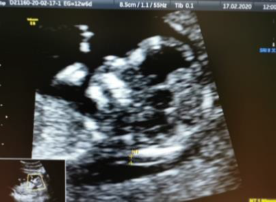
In the maternal-fetal consultation, a first-trimester ultrasound is performed where screening for chromopathies is performed through nuchal translucency (Figure 1) and second markers, which are within normal limits, with risks of 1 in 5,000 for trisomy 21, 1 in 5,000 for trisomy 18; 1 in 10,000 for trisomy 13, remaining in the low risk group.

Figure 1 The maternal-fetal consultation, a first-trimester ultrasound is performed where screening for chromopathies is performed through nuchal translucency.
In the placental area, a snowflake image is displayed (Figure 2).
Quantification of human chorionic gonadotropin was requested, which was initially reported at values of 180,944.77mIUml, 48 hours later, a new quantification was requested, which was reported with high values of 225,464mIUml. The patient is informed of the need to perform an invasive study to perform a karyotype, however the patient does not accept due to the risk of losing the pregnancy, prenatal control is continued as usual in the maternal-fetal service where prophylactic management is given to hypertensive disease with acetylsalicylic acid 150mg every 24 hours at night and complementary laboratory studies are requested for trophoblastic disease blood cytometry with hemoglobin 13.9mg/dl, leukocytes 10.9 x3, platelets 278 thousand, Rh + group, glucose 75mg/dl. BUN 6.0 Creatinine 0.56, total bile 0.83, TGP 14 TGO 21, DHL 181 PT 12.7 TTp 28 INR 0.97, non-pathological general urine test, chest X-ray and liver ultrasound imaging studies without alterations, an open appointment is given to the emergency room with alarm data and appointment in 4 weeks.
On this occasion, already at 17 weeks of gestation, an ultrasound is performed in which a single live fetus is visualized, with an estimated fetal weight of 181g, qualitatively normal amniotic fluid, with posterior corporeal placenta with multiple vesicles which give an image compatible with an incomplete mole. brings with it quantification of which was reported in 243 188.96mUml. An appointment is made to perform a structural ultrasound at week 23 of gestation, where the fetus is reported without anatomical alterations, with ammonia growth according to the gestational age data, with the following measurements:
Parameter |
Measurement (CM) |
DBP |
5.12 CM |
CC |
19.88 CM |
CA |
18.92 CM |
LF |
4.13 CM |
CUBITO |
3.38 CM |
TIB |
3.66 CM |
RAD |
2.95 CM |
PER |
3.52 CM |
FCF |
142 LPM |
ESTIMATED WEIGHT |
581 GR |
ACM |
IP 2.14 |
AUL |
IP 1.07 |
AUR |
IP 0.72 |
AU |
IP 1.0 |
Prenatal control was continued by the maternal-fetal medicine service without reporting alterations in subsequent ultrasounds, monitoring of human chorionic gonatropin concentrations continued, with a significant decrease reported for April 14, 2020; 13 600.10mIUml, rest of paraclinical within normal parameters, likewise thyroid profile with TSH 2.10, total T3 of 1.68ng/ml total T4 of 1.37.
At week 36 of gestation, a new quantification of human chorionic gonatropin was requested, which was reported on this occasion at 3,170mUIml, the rest of the laboratories without alterations. Reason for which it was decided to schedule an elective caesarean section on August 4, 2020, obtaining a female newborn weighing 2730 gr Size 45cm Apgar 8–9, Capurro 38 SDG, Silverman 0, with birth time of 08:50hrs, reporting a bleeding of 250ml, visualizing multiple cystic formations in the placenta, after anesthetic recovery, she went up to the obstetrics hospitalization area, where she evolved favorably, and the mother and daughter were discharged from the service without complications.
The pathology service report reports a placental disc of 20x18x3cm, a central cord of 23x1cm, weighing 780g. The opaque light brown membranous fetal side, the maternal side lobed wine red with a hemorrhagic appearance; 17 cotyledons are identified, on the fetal face the vessels are congestive tortuous, trivascular umbilical cord. On section, multiple cystic lesions of approximately 0.5cm and axis of hyaline content are identified. Concluding in the diagnosis placenta of third trimester of gestation, fragmented and deformed, monochorionic, amniotic. gestational trophoblastic disease, partial mole, chorionic villi with hydropic villous dilatation, calcification villous dystrophy (Figure 3 & 4).
Partial Hydatidiform Mole (MHP): The karyotype is generally triploid (69XYY or 69XXY), which can be produced by 3 mechanisms:
Characteristics of a normally developing placenta and a complete hydatidiform mole occur concurrently, with a range of villi from normal to cystic, whereas trophoblast hyperplasia is only focal or “patchy” and usually involves the syncytiotrophoblast.
In some cases of MHP the fetus is present, but its development is almost always abnormal, due to associated chromosomal abnormalities (triploidy). Clinical presentation:
It is common for the clinical picture to be the manifestations of an ongoing or incomplete abortion.
Gynaecorrhagia is present in 72% of patients.
Uterine height higher than expected for gestational age (3.7%).
Preeclampsia (2.5%).
Low association with hyperthyroidism, hyperemesis gravidarum and theco-lutein cysts. This entity behaves benignly in most cases and the risk of malignant transformation is around 4%.
The coexistence of molar degeneration of the placenta and fetus in the same pregnancy is an infrequent fact that can occur in 0.005-1% of all pregnancies, this being even rarer in multiple gestations, even so there are several cases described in the medical literature due to the persistence of gestational trophoblastic disease in complete moles. When a fetus coexists with hydatidiform degeneration of the placenta, the difference between complete and incomplete mole is of relevant interest to assess the aggressiveness and prognosis of said disease, which is why incomplete moles must be differentiated from dizygotic twin pregnancies. For complete mole and normal fetus.3,4
Incomplete mole is diagnosed based on ultrasound findings, a partial mole is diagnosed as a missed or incomplete abortion in 15 to 60 percent of cases. These misdiagnoses are more common in partial mole because they are accompanied by a fetus and amniotic fluid. Sonographic features suggestive of a partial molar pregnancy include:
The existence of a live fetus is a rare case that occurs in 1 in 22,000 pregnancies or 1 in 100,000. In this case, it is an exceptional case, of which there are very few reported in the medical literature.
None.
None.
The authors declare having no conflict of interest.

©2022 , et al. This is an open access article distributed under the terms of the, which permits unrestricted use, distribution, and build upon your work non-commercially.